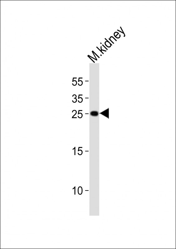
Western blot analysis of lysate from mouse kidney tissue lysate, using Cldn6 Antibody (C-term). M08182-1 was diluted at 1:1000. A goat anti-rabbit IgG H&L (HRP) at 1:10000 dilution was used as the secondary antibody. Lysate at 20ug.
all(2)
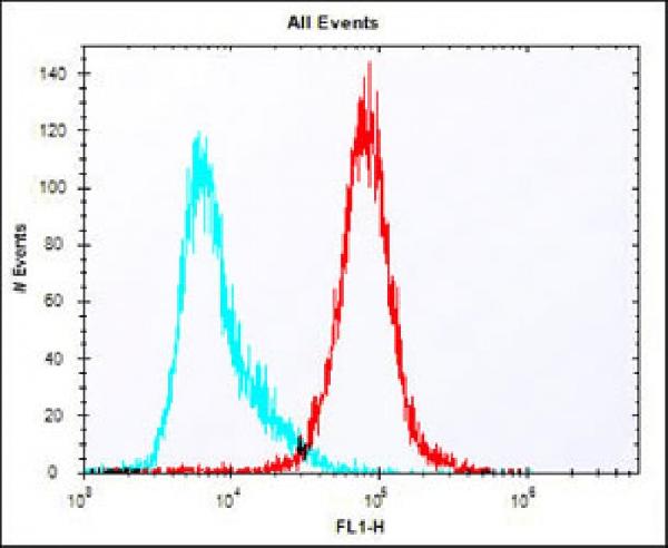
Overlay histogram showing colon26 cells stained with M08182-1 (red line). The cells were fixed with 2% paraformaldehyde (10 min) and then permeabilized with 90% methanol for 10 min. The cells were then icubated in 2% bovine serum albumin to block non-specific protein-protein interactions followed by the antibody (M08182-1, 1:25 dilution) for 60 min at 37oC. The secondary antibody used was Alexa Fluor? 488 goat anti-rabbit lgG (H+L) at 1/400 dilution for 40 min at 37oC. Isotype control antibody (blue line) was rabbit IgG1 (1μg/1x10^6 cells) used under the same conditions. Acquisition of >10, 000 events was performed.
all(2) Western blot analysis of lysate from mouse kidney tissue lysate, using Cldn6 Antibody (C-term). M08182-1 was diluted at 1:1000. A goat anti-rabbit IgG H&L (HRP) at 1:10000 dilution was used as the secondary antibody. Lysate at 20ug.
Overlay histogram showing colon26 cells stained with M08182-1 (red line). The cells were fixed with 2% paraformaldehyde (10 min) and then permeabilized with 90% methanol for 10 min. The cells were then icubated in 2% bovine serum albumin to block non-specific protein-protein interactions followed by the antibody (M08182-1, 1:25 dilution) for 60 min at 37oC. The secondary antibody used was Alexa Fluor? 488 goat anti-rabbit lgG (H+L) at 1/400 dilution for 40 min at 37oC. Isotype control antibody (blue line) was rabbit IgG1 (1μg/1x10^6 cells) used under the same conditions. Acquisition of >10, 000 events was performed.
 联系我们
联系我们800-880-8748
 关注我们
关注我们
本司产品仅用于科研,不用于临床诊断和治疗
联系方式:027-67845390 8008808748 技术支持:武汉丰网
© 1993-2024 Boster Biological Technology Co.,Ltd E-mail:boster@boster.com 鄂ICP备05005548号-1  鄂公网安备 42018502007312号

 积分商城
积分商城  购物车
购物车  登录/注册
登录/注册  您当前的位置:
  说明书
说明书


 成功添加到购物车
 微信客服
微信客服


 微信扫一扫立即咨询