| Western blot (WB): | 1:2000 |
| Immunohistochemistry (IHC): | 1:50 |
| Immunocytochemistry/Immunofluorescence (ICC/IF): | 1:100 |
| Flow Cytometry(FCM): | 1:100 |
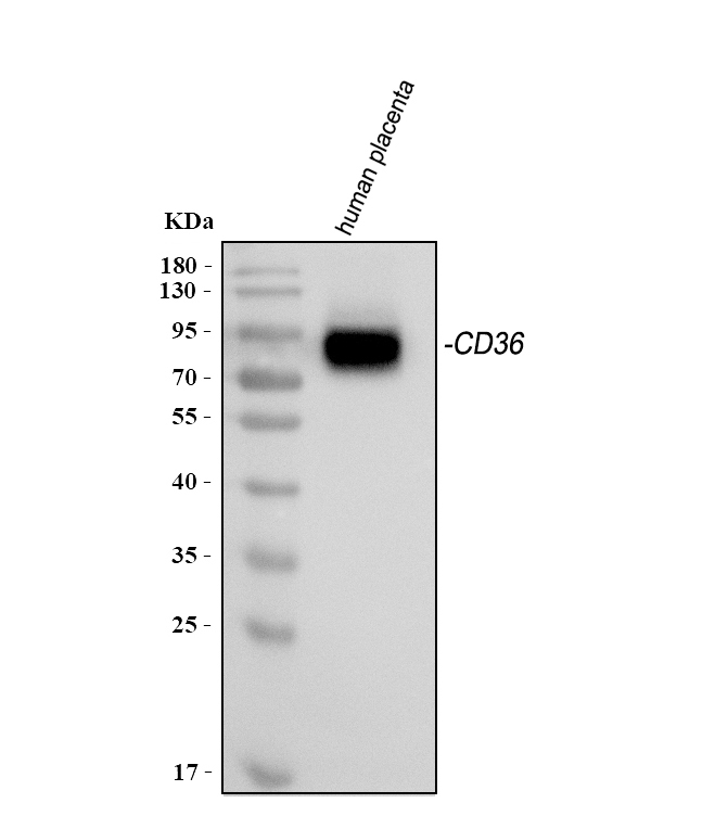
Western blot analysis of anti-CD36 antibody (MA01189). The sample well of each lane was loaded with 30 ug of sample under reducing conditions.
Lane 1: human placenta tissue lysates.
After electrophoresis, proteins were transferred to a membrane. Then the membrane was incubated with mouse anti-CD36 antigen affinity purified monoclonal antibody (MA01189) and probed with a goat anti-mouse IgG-HRP secondary antibody (Catalog # BA1050). The signal is developed using ECL Plus Western Blotting Substrate (Catalog # AR1197). A specific band was detected for CD36 at approximately 88 kDa. The expected band size for CD36 is at 53 kDa.
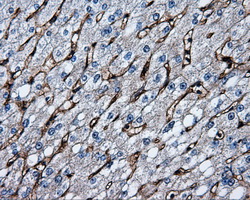
Immunohistochemical staining of paraffin-embedded liver tissue within the normal limits using anti-CD36mouse monoclonal antibody. (Heat-induced epitope retrieval by 10mM citric buffer, pH6.0, 100°C for 10min, M01189-1, Dilution 1:50)
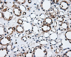
Immunohistochemical staining of paraffin-embedded Kidney tissue within the normal limits using anti-CD36mouse monoclonal antibody. (Heat-induced epitope retrieval by 10mM citric buffer, pH6.0, 100°C for 10min, M01189-1, Dilution 1:50)
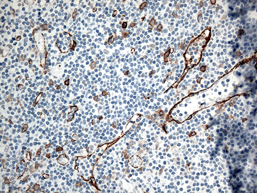
Immunohistochemical staining of paraffin-embedded Human tonsil within the normal limits using anti-CD36 mouse monoclonal antibody. (Heat-induced epitope retrieval by 1mM EDTA in 10mM Tris buffer (pH8.5) at 120°C for 3min, M01189-1) (1:500)
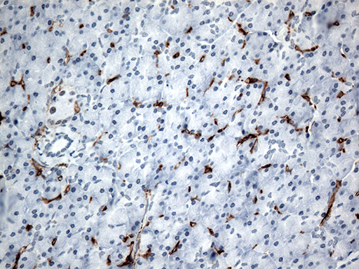
Immunohistochemical staining of paraffin-embedded Human pancreas tissue within the normal limits using anti-CD36 mouse monoclonal antibody. (Heat-induced epitope retrieval by 1mM EDTA in 10mM Tris buffer (pH8.5) at 120°C for 3min, M01189-1) (1:500)
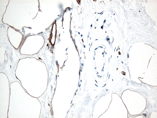
Immunohistochemical staining of paraffin-embedded Human breast tissue within the normal limits using anti-CD36 mouse monoclonal antibody. (Heat-induced epitope retrieval by 1mM EDTA in 10mM Tris buffer (pH8.5) at 120°C for 3min, M01189-1) (1:500)
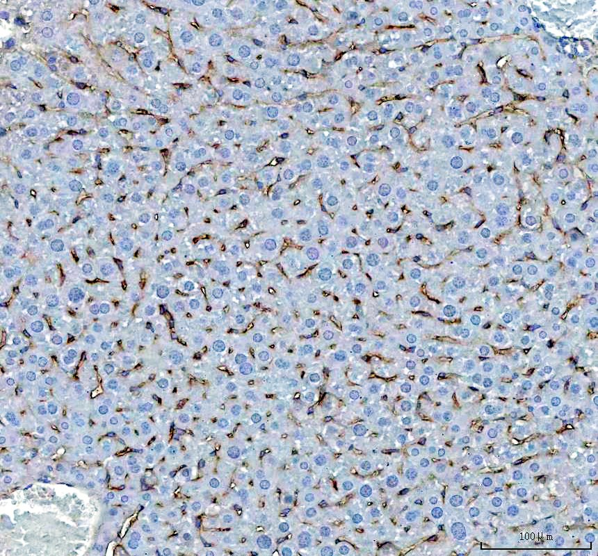
IHC analysis of CD36 using anti-CD36 antibody (MA01189).
CD36 was detected in a paraffin-embedded section of mouse liver tissue. The tissue section was incubated with mouse anti-CD36 Antibody (MA01189) at a dilution of 1:200 and developed using HRP Conjugated mouse IgG Super Vision Assay Kit (Catalog # SV0001) with DAB (Catalog # AR1027) as the chromogen.
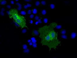
Anti-CD36 mouse monoclonal antibody immunofluorescent staining of COS7 cells transiently transfected by pCMV6-ENTRY CD36.
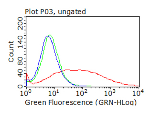
HEK293T cells transfected with either overexpress plasmid (Red), compared to an IgG isotype control, (Green) or empty vector control plasmid (Blue) were immunostained by anti-CD36 antibody, and then analyzed by flow cytometry (1:100).

Western blot analysis of anti-CD36 antibody (MA01189). The sample well of each lane was loaded with 30 ug of sample under reducing conditions.
Lane 1: human placenta tissue lysates.
After electrophoresis, proteins were transferred to a membrane. Then the membrane was incubated with mouse anti-CD36 antigen affinity purified monoclonal antibody (MA01189) and probed with a goat anti-mouse IgG-HRP secondary antibody (Catalog # BA1050). The signal is developed using ECL Plus Western Blotting Substrate (Catalog # AR1197). A specific band was detected for CD36 at approximately 88 kDa. The expected band size for CD36 is at 53 kDa.

Immunohistochemical staining of paraffin-embedded liver tissue within the normal limits using anti-CD36mouse monoclonal antibody. (Heat-induced epitope retrieval by 10mM citric buffer, pH6.0, 100°C for 10min, M01189-1, Dilution 1:50)

Immunohistochemical staining of paraffin-embedded Kidney tissue within the normal limits using anti-CD36mouse monoclonal antibody. (Heat-induced epitope retrieval by 10mM citric buffer, pH6.0, 100°C for 10min, M01189-1, Dilution 1:50)

Immunohistochemical staining of paraffin-embedded Human tonsil within the normal limits using anti-CD36 mouse monoclonal antibody. (Heat-induced epitope retrieval by 1mM EDTA in 10mM Tris buffer (pH8.5) at 120°C for 3min, M01189-1) (1:500)

Immunohistochemical staining of paraffin-embedded Human pancreas tissue within the normal limits using anti-CD36 mouse monoclonal antibody. (Heat-induced epitope retrieval by 1mM EDTA in 10mM Tris buffer (pH8.5) at 120°C for 3min, M01189-1) (1:500)

Immunohistochemical staining of paraffin-embedded Human breast tissue within the normal limits using anti-CD36 mouse monoclonal antibody. (Heat-induced epitope retrieval by 1mM EDTA in 10mM Tris buffer (pH8.5) at 120°C for 3min, M01189-1) (1:500)

IHC analysis of CD36 using anti-CD36 antibody (MA01189).
CD36 was detected in a paraffin-embedded section of mouse liver tissue. The tissue section was incubated with mouse anti-CD36 Antibody (MA01189) at a dilution of 1:200 and developed using HRP Conjugated mouse IgG Super Vision Assay Kit (Catalog # SV0001) with DAB (Catalog # AR1027) as the chromogen.

Anti-CD36 mouse monoclonal antibody immunofluorescent staining of COS7 cells transiently transfected by pCMV6-ENTRY CD36.

HEK293T cells transfected with either overexpress plasmid (Red), compared to an IgG isotype control, (Green) or empty vector control plasmid (Blue) were immunostained by anti-CD36 antibody, and then analyzed by flow cytometry (1:100).


