| Western blot (WB): | 1:2000 |
| Immunocytochemistry/Immunofluorescence (ICC/IF): | 1:100 |
| Flow cytometry (FCM): | 1:100 |
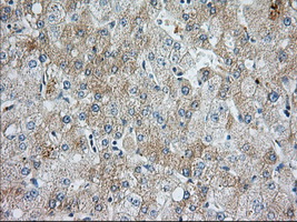
Immunohistochemical staining of paraffin-embedded Human liver tissue within the normal limits using anti-BIRC5 mouse monoclonal antibody. (Heat-induced epitope retrieval by 1mM EDTA in 10mM Tris, pH9.0, 120°C for 3min, MA00379, Dilution 1:50)
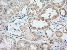
Immunohistochemical staining of paraffin-embedded Human Kidney tissue within the normal limits using anti-BIRC5 mouse monoclonal antibody. (Heat-induced epitope retrieval by 1mM EDTA in 10mM Tris, pH9.0, 120°C for 3min, MA00379, Dilution 1:50)
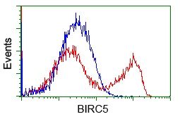
HEK293T cells transfected with either overexpress plasmid (Red) or empty vector control plasmid (Blue) were immunostained by anti-BIRC5 antibody, and then analyzed by flow cytometry.
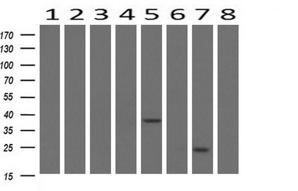
Western blot analysis of extracts (10ug) from 8 Human tissue by using anti-BIRC5 monoclonal antibody at 1:200 (1: Testis; 2: Uterus; 3: Breast; 4: Brain; 5: Liver; 6: Ovary; 7: Thyroid gland; 8: Colon).
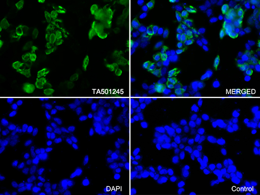
Immunofluorescent staining of 293T cells transfected by pCMV6-ENTRY BIRC5 using anti-BIRC5 antibody. 293T cells transfected with empty vector served as a negative control (MERGED, lower right) (1:100).
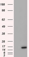
HEK293T cells were transfected with the pCMV6-ENTRY control (Left lane) or pCMV6-ENTRY BIRC5 (Right lane) cDNA for 48 hrs and lysed. Equivalent amounts of cell lysates (5 ug per lane) were separated by SDS-PAGE and immunoblotted with anti-BIRC5 (Cat# MA00379).

Immunohistochemical staining of paraffin-embedded Human liver tissue within the normal limits using anti-BIRC5 mouse monoclonal antibody. (Heat-induced epitope retrieval by 1mM EDTA in 10mM Tris, pH9.0, 120°C for 3min, MA00379, Dilution 1:50)

Immunohistochemical staining of paraffin-embedded Human Kidney tissue within the normal limits using anti-BIRC5 mouse monoclonal antibody. (Heat-induced epitope retrieval by 1mM EDTA in 10mM Tris, pH9.0, 120°C for 3min, MA00379, Dilution 1:50)

HEK293T cells transfected with either overexpress plasmid (Red) or empty vector control plasmid (Blue) were immunostained by anti-BIRC5 antibody, and then analyzed by flow cytometry.

Western blot analysis of extracts (10ug) from 8 Human tissue by using anti-BIRC5 monoclonal antibody at 1:200 (1: Testis; 2: Uterus; 3: Breast; 4: Brain; 5: Liver; 6: Ovary; 7: Thyroid gland; 8: Colon).

Immunofluorescent staining of 293T cells transfected by pCMV6-ENTRY BIRC5 using anti-BIRC5 antibody. 293T cells transfected with empty vector served as a negative control (MERGED, lower right) (1:100).

HEK293T cells were transfected with the pCMV6-ENTRY control (Left lane) or pCMV6-ENTRY BIRC5 (Right lane) cDNA for 48 hrs and lysed. Equivalent amounts of cell lysates (5 ug per lane) were separated by SDS-PAGE and immunoblotted with anti-BIRC5 (Cat# MA00379).


