| Western blot (WB): | 1:500-2000 |

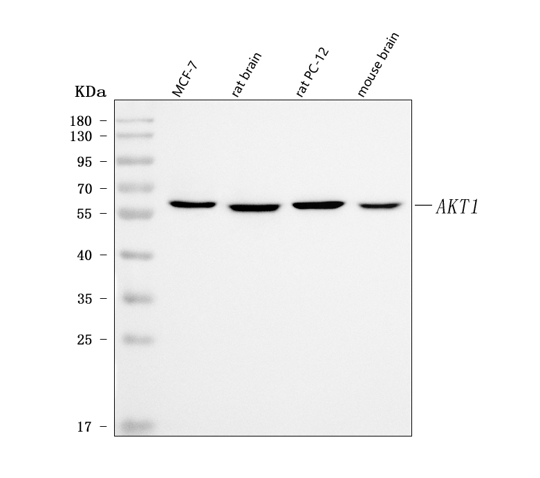
Western blot analysis of anti- AKT1 antibody (BM1612). The sample well of each lane was loaded with 30ug of sample under reducing conditions.
Lane 1: human MCF-7 whole cell lysates,
Lane 2: rat brain tissue lysates,
Lane 3: rat PC-12 whole cell lysates,
Lane 4: mouse brain tissue lysates.
Use mouse anti- AKT1 1:1000, probed with a goat anti- mouse IgG-HRP secondary antibody. The signal is developed using an Enhanced Chemiluminescent detection (ECL) kit (Catalog#EK1001). A specific band was detected for AKT1 at approximately 60KD. The expected band size for AKT1 is at 60KD.
