| Western blot (WB): | 1:500~2000 |
| Immunohistochemistry (IHC): | 1:150 |
| Immunocytochemistry/Immunofluorescence (ICC/IF): | 1:100 |
| Flow cytometry (FCM): | 1:100 |
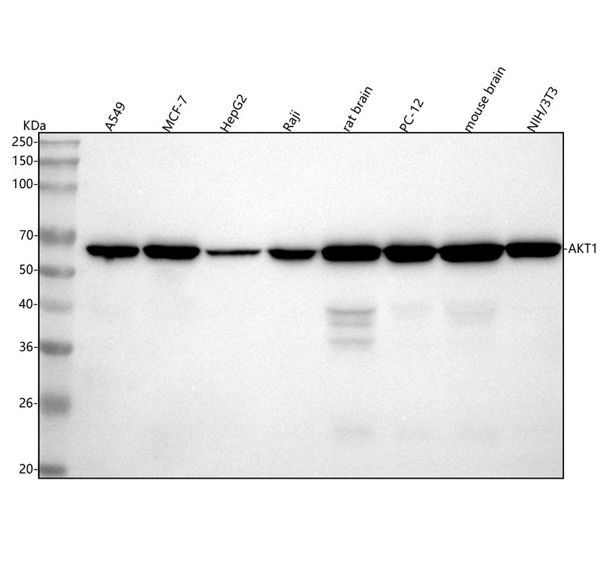
Western blot analysis of anti-AKT1 antibody (M00024). The sample well of each lane was loaded with 30 ug of sample under reducing conditions.
Lane 1: human A549 whole cell lysates,
Lane 2: human MCF-7 whole cell lysates,
Lane 3: human HepG2 whole cell lysates,
Lane 4: human Raji whole cell lysates,
Lane 5: rat brain tissue lysates,
Lane 6: rat PC-12 whole cell lysates,
Lane 7: mouse brain tissue lysates,
Lane 8: mouse NIH/3T3 whole cell lysates.
After electrophoresis, proteins were transferred to a membrane. Then the membrane was incubated with mouse anti-AKT1 antigen affinity purified monoclonal antibody (M00024) at a dilution of 1:1000 and probed with a goat anti-mouse IgG-HRP secondary antibody (Catalog # BA1050). The signal is developed using ECL Plus Western Blotting Substrate (Catalog # AR1197). A specific band was detected for AKT1 at approximately 56 kDa. The expected band size for AKT1 is at 56 kDa.
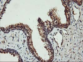
Immunohistochemical staining of paraffin-embedded Human prostate tissue within the normal limits using anti-AKT1 mouse monoclonal antibody. (Heat-induced epitope retrieval by 10mM citric buffer, pH6.0, 100°C for 10min, M00024)
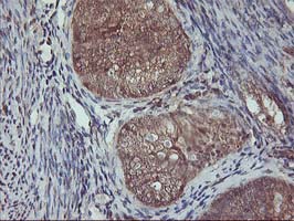
Immunohistochemical staining of paraffin-embedded Adenocarcinoma of Human endometrium tissue using anti-AKT1 mouse monoclonal antibody. (Heat-induced epitope retrieval by 10mM citric buffer, pH6.0, 100°C for 10min, M00024)
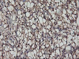
Immunohistochemical staining of paraffin-embedded Carcinoma of Human kidney tissue using anti-AKT1 mouse monoclonal antibody. (Heat-induced epitope retrieval by 10mM citric buffer, pH6.0, 100°C for 10min, M00024)
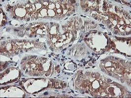
Immunohistochemical staining of paraffin-embedded Human Kidney tissue within the normal limits using anti-AKT1 mouse monoclonal antibody. (Heat-induced epitope retrieval by 10mM citric buffer, pH6.0, 100°C for 10min, M00024)
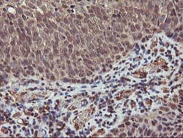
Immunohistochemical staining of paraffin-embedded Carcinoma of Human bladder tissue using anti-AKT1 mouse monoclonal antibody. (Heat-induced epitope retrieval by 10mM citric buffer, pH6.0, 100°C for 10min, M00024)
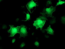
Anti-AKT1 mouse monoclonal antibody immunofluorescent staining of COS7 cells transiently transfected by pCMV6-ENTRY AKT1 .

Flow cytometric Analysis of Jurkat cells, using anti-AKT1 antibody, (Red), compared to a nonspecific negative control antibody, (Blue).

Western blot analysis of anti-AKT1 antibody (M00024). The sample well of each lane was loaded with 30 ug of sample under reducing conditions.
Lane 1: human A549 whole cell lysates,
Lane 2: human MCF-7 whole cell lysates,
Lane 3: human HepG2 whole cell lysates,
Lane 4: human Raji whole cell lysates,
Lane 5: rat brain tissue lysates,
Lane 6: rat PC-12 whole cell lysates,
Lane 7: mouse brain tissue lysates,
Lane 8: mouse NIH/3T3 whole cell lysates.
After electrophoresis, proteins were transferred to a membrane. Then the membrane was incubated with mouse anti-AKT1 antigen affinity purified monoclonal antibody (M00024) at a dilution of 1:1000 and probed with a goat anti-mouse IgG-HRP secondary antibody (Catalog # BA1050). The signal is developed using ECL Plus Western Blotting Substrate (Catalog # AR1197). A specific band was detected for AKT1 at approximately 56 kDa. The expected band size for AKT1 is at 56 kDa.

Immunohistochemical staining of paraffin-embedded Human prostate tissue within the normal limits using anti-AKT1 mouse monoclonal antibody. (Heat-induced epitope retrieval by 10mM citric buffer, pH6.0, 100°C for 10min, M00024)

Immunohistochemical staining of paraffin-embedded Adenocarcinoma of Human endometrium tissue using anti-AKT1 mouse monoclonal antibody. (Heat-induced epitope retrieval by 10mM citric buffer, pH6.0, 100°C for 10min, M00024)

Immunohistochemical staining of paraffin-embedded Carcinoma of Human kidney tissue using anti-AKT1 mouse monoclonal antibody. (Heat-induced epitope retrieval by 10mM citric buffer, pH6.0, 100°C for 10min, M00024)

Immunohistochemical staining of paraffin-embedded Human Kidney tissue within the normal limits using anti-AKT1 mouse monoclonal antibody. (Heat-induced epitope retrieval by 10mM citric buffer, pH6.0, 100°C for 10min, M00024)

Immunohistochemical staining of paraffin-embedded Carcinoma of Human bladder tissue using anti-AKT1 mouse monoclonal antibody. (Heat-induced epitope retrieval by 10mM citric buffer, pH6.0, 100°C for 10min, M00024)

Anti-AKT1 mouse monoclonal antibody immunofluorescent staining of COS7 cells transiently transfected by pCMV6-ENTRY AKT1 .

Flow cytometric Analysis of Jurkat cells, using anti-AKT1 antibody, (Red), compared to a nonspecific negative control antibody, (Blue).


