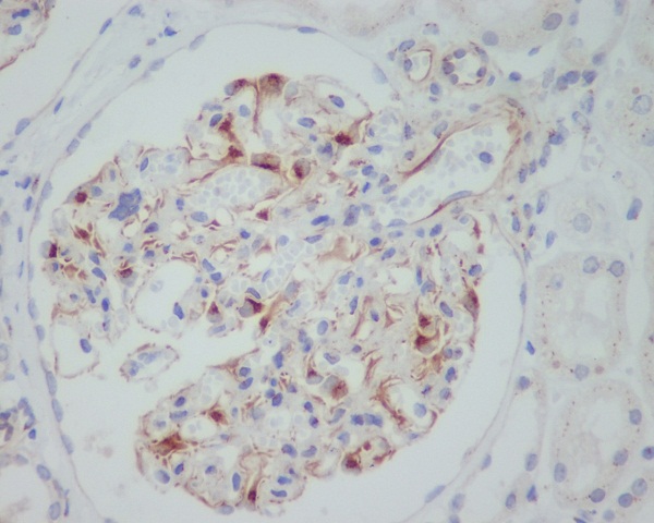
Immunohistochemical staining of paraffin-embedded Carcinoma of Human liver tissue using anti-VEGF mouse monoclonal antibody. (MA00045)
all(6)
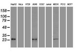
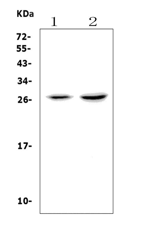
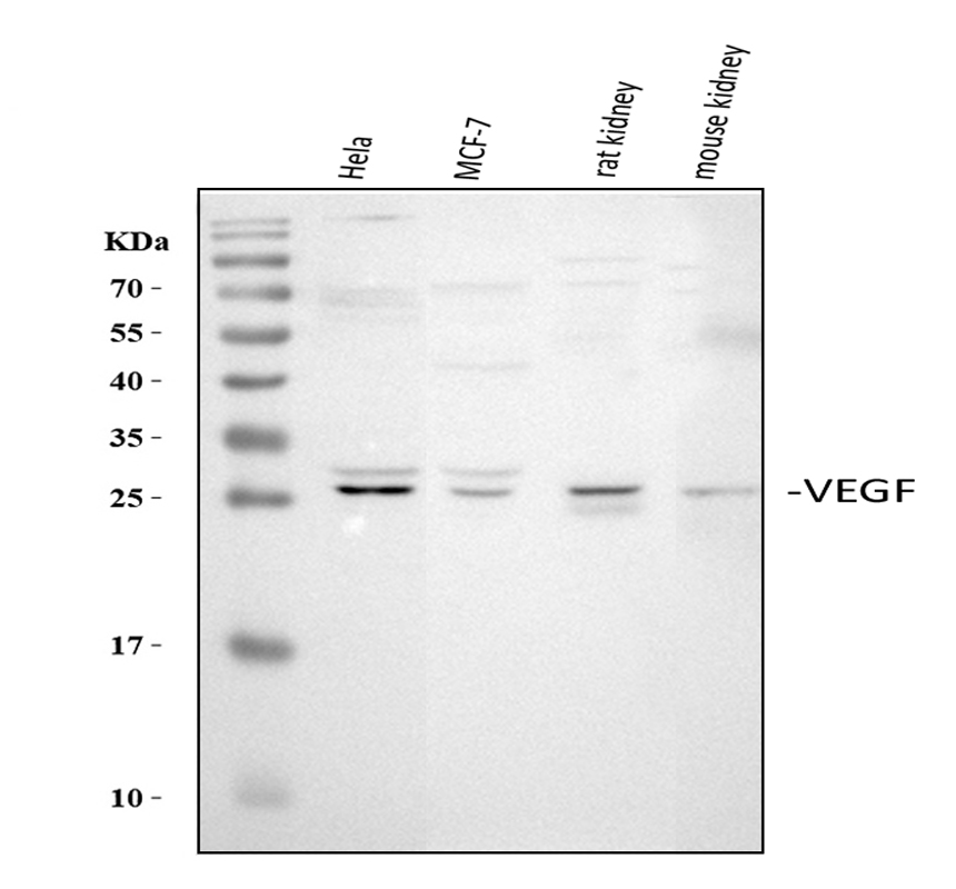
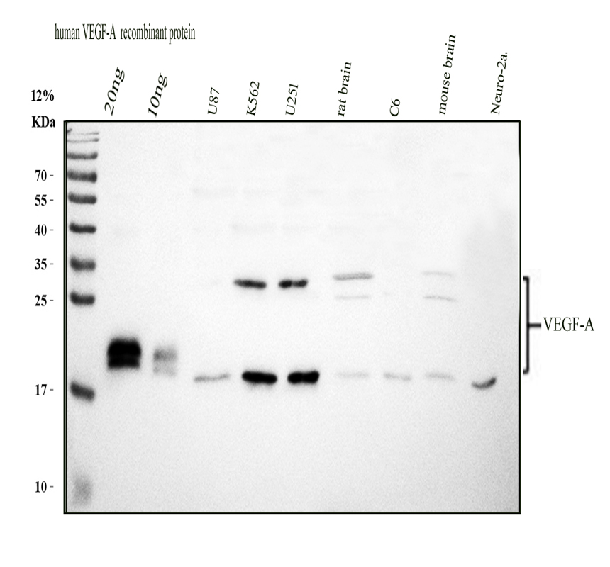
Western blot analysis of extracts (35ug) from 9 different cell lines by using anti-VEGF monoclonal antibody.
all(6) 
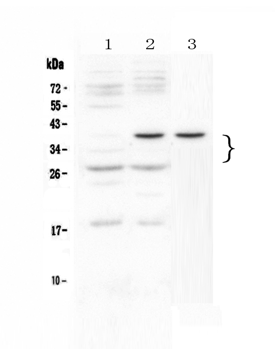
HEK293T cells were transfected with the overexpression plasmids of 9 VEGF isoforms (from left to right:VEGF206, ; VEGF189, ; L-VEGF165, ; VEGF165, ; L-VEGF121, ; VEGF iso18, ; VEGF183, ; VEGF145, ; L-VEGF189,) for 48 hrs and lysed. Equivalent amounts of cell lysates (5 ug per lane) were separated by SDS-PAGE and immunoblotted with anti-flag antibody (1:1000) or anti-VEGFA mouse monoclonal antibody. (MA00045, 1:500)
all(6) 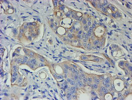
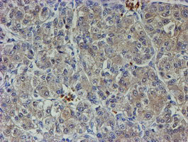
Immunohistochemical staining of paraffin-embedded Carcinoma of Human pancreas tissue using anti-VEGF mouse monoclonal antibody. (MA00045)
all(6) 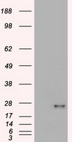
HEK293T cells were transfected with the pCMV6-ENTRY control (Left lane) or pCMV6-ENTRY VEGF (Right lane) cDNA for 48 hrs and lysed. Equivalent amounts of cell lysates (5 ug per lane) were separated by SDS-PAGE and immunoblotted with anti-VEGF.
all(6) 
| Western blot (WB): | 1:500-2000 |
| Immunohistochemistry (IHC): | 1:50-400 |
| Immunocytochemistry/Immunofluorescence (ICC/IF): | 1:50-400 |
Immunohistochemical staining of paraffin-embedded Carcinoma of Human liver tissue using anti-VEGF mouse monoclonal antibody. (MA00045)
Western blot analysis of extracts (35ug) from 9 different cell lines by using anti-VEGF monoclonal antibody.
HEK293T cells were transfected with the overexpression plasmids of 9 VEGF isoforms (from left to right:VEGF206, ; VEGF189, ; L-VEGF165, ; VEGF165, ; L-VEGF121, ; VEGF iso18, ; VEGF183, ; VEGF145, ; L-VEGF189,) for 48 hrs and lysed. Equivalent amounts of cell lysates (5 ug per lane) were separated by SDS-PAGE and immunoblotted with anti-flag antibody (1:1000) or anti-VEGFA mouse monoclonal antibody. (MA00045, 1:500)
Immunohistochemical staining of paraffin-embedded Carcinoma of Human pancreas tissue using anti-VEGF mouse monoclonal antibody. (MA00045)
HEK293T cells were transfected with the pCMV6-ENTRY control (Left lane) or pCMV6-ENTRY VEGF (Right lane) cDNA for 48 hrs and lysed. Equivalent amounts of cell lysates (5 ug per lane) were separated by SDS-PAGE and immunoblotted with anti-VEGF.
 联系我们
联系我们027-67845390
 关注我们
关注我们
本司产品仅用于科研,不用于临床诊断和治疗
联系方式:027-67845390/1/2 技术支持:武汉丰网
© 1993-2025 Boster Biological Technology co.Itd E-mail:boster@boster.com
鄂ICP备05005548号-2
 鄂公网安备 42018502007312号

 积分商城
积分商城  购物车
购物车  登录/注册
登录/注册  您当前的位置:
  说明书
说明书 一键复制产品信息
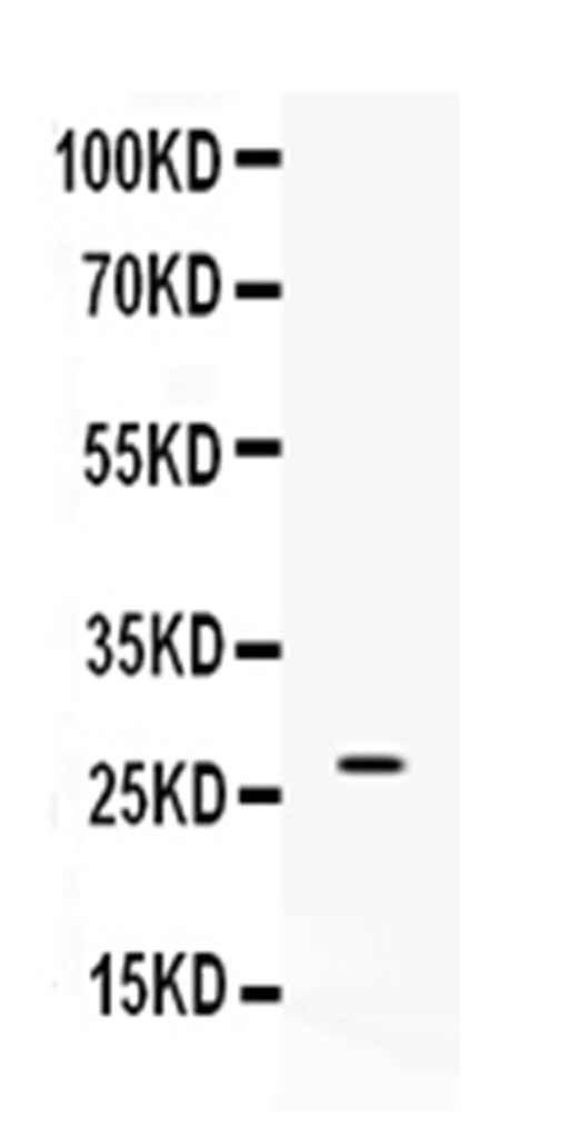
 成功添加到购物车
 微信客服
微信客服


 微信扫一扫立即咨询
