| Western blot (WB): | 1:500-2000 |
| Immunohistochemistry (IHC): | 1:50-400 |
| Enzyme linked immunosorbent assay (ELISA): | 1:100-1000 |
| (Boiling the paraffin sections in 10mM citrate buffer,pH6.0,or PH8.0 EDTA repair liquid for 20 mins is required for the staining of formalin/paraffin sections.) Optimal working dilutions must be determined by end user. | |

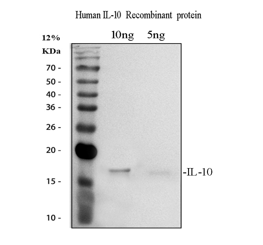
Western blot analysis of IL10 using anti-IL10 antibody (RP1014).
Lane 1: recombinant human IL10 protein 10 ng,
Lane 2: recombinant human IL10 protein 5 ng.
After electrophoresis, proteins were transferred to a membrane. Then the membrane was incubated with rabbit anti-IL10 antigen affinity purified polyclonal antibody (RP1014) at a dilution of 1:1000 and probed with a goat anti-rabbit IgG-HRP secondary antibody (Catalog # BA1054). The signal is developed using ECL Plus Western Blotting Substrate (Catalog # AR1197). A specific band was detected for IL10 at approximately 17 kDa.

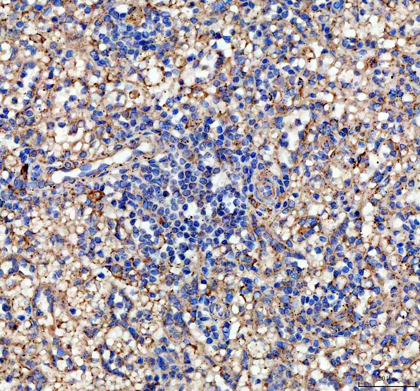
IHC analysis of IL10 using anti-IL10 antibody (RP1014).
IL10 was detected in a paraffin-embedded section of human spleen tissue. The tissue section was incubated with rabbit anti-IL10 Antibody (RP1014) at a dilution of 1:200 and developed using HRP Conjugated Rabbit IgG Super Vision Assay Kit (Catalog # SV0002) with DAB (Catalog # AR1027) as the chromogen.

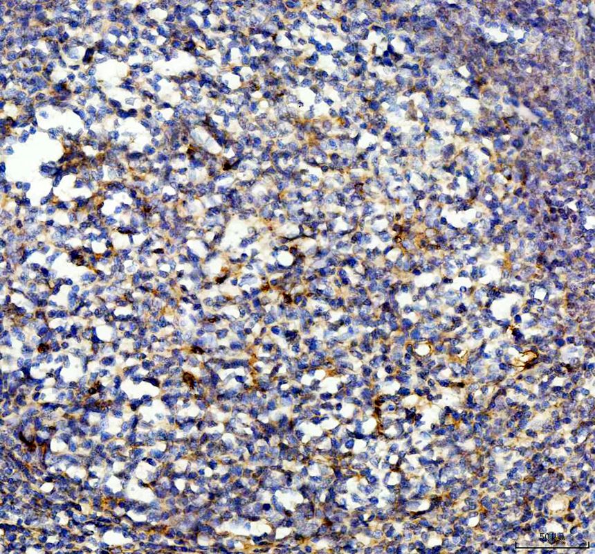
IHC analysis of IL10 using anti-IL10 antibody (RP1014).
IL10 was detected in a paraffin-embedded section of human appendix tissue. The tissue section was incubated with rabbit anti-IL10 Antibody (RP1014) at a dilution of 1:200 and developed using HRP Conjugated Rabbit IgG Super Vision Assay Kit (Catalog # SV0002) with DAB (Catalog # AR1027) as the chromogen.

Western blot analysis of IL10 using anti-IL10 antibody (RP1014).
Lane 1: recombinant human IL10 protein 10 ng,
Lane 2: recombinant human IL10 protein 5 ng.
After electrophoresis, proteins were transferred to a membrane. Then the membrane was incubated with rabbit anti-IL10 antigen affinity purified polyclonal antibody (RP1014) at a dilution of 1:1000 and probed with a goat anti-rabbit IgG-HRP secondary antibody (Catalog # BA1054). The signal is developed using ECL Plus Western Blotting Substrate (Catalog # AR1197). A specific band was detected for IL10 at approximately 17 kDa.

IHC analysis of IL10 using anti-IL10 antibody (RP1014).
IL10 was detected in a paraffin-embedded section of human spleen tissue. The tissue section was incubated with rabbit anti-IL10 Antibody (RP1014) at a dilution of 1:200 and developed using HRP Conjugated Rabbit IgG Super Vision Assay Kit (Catalog # SV0002) with DAB (Catalog # AR1027) as the chromogen.

IHC analysis of IL10 using anti-IL10 antibody (RP1014).
IL10 was detected in a paraffin-embedded section of human appendix tissue. The tissue section was incubated with rabbit anti-IL10 Antibody (RP1014) at a dilution of 1:200 and developed using HRP Conjugated Rabbit IgG Super Vision Assay Kit (Catalog # SV0002) with DAB (Catalog # AR1027) as the chromogen.


