
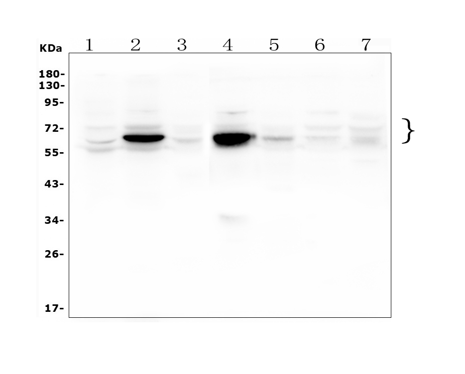
Western blot analysis of Beta Galactosidase/GLB1 using anti-Beta Galactosidase/GLB1 antibody (A01829-3). The sample well of each lane was loaded with 30 ug of sample under reducing conditions.
Lane 1: rat lung tissue lysates,
Lane 2: rat liver tissue lysates,
Lane 3: rat testis tissue lysates,
Lane 4: mouse liver tissue lysates,
Lane 5: mouse testis tissue lysates,
Lane 6: mouse HEPA1-6 whole cell lysates,
Lane 7: mouse Neuro-2a whole cell lysates.
After electrophoresis, proteins were transferred to a membrane. Then the membrane was incubated with rabbit anti-Beta Galactosidase/GLB1 antigen affinity purified polyclonal antibody (A01829-3) at a dilution of 1:1000 and probed with a goat anti-rabbit IgG-HRP secondary antibody (Catalog # BA1054). The signal is developed using ECL Plus Western Blotting Substrate (Catalog # AR1197). A specific band was detected for Beta Galactosidase/GLB1 at approximately 65-85 kDa. The expected band size for Beta Galactosidase/GLB1 is at 73 kDa.

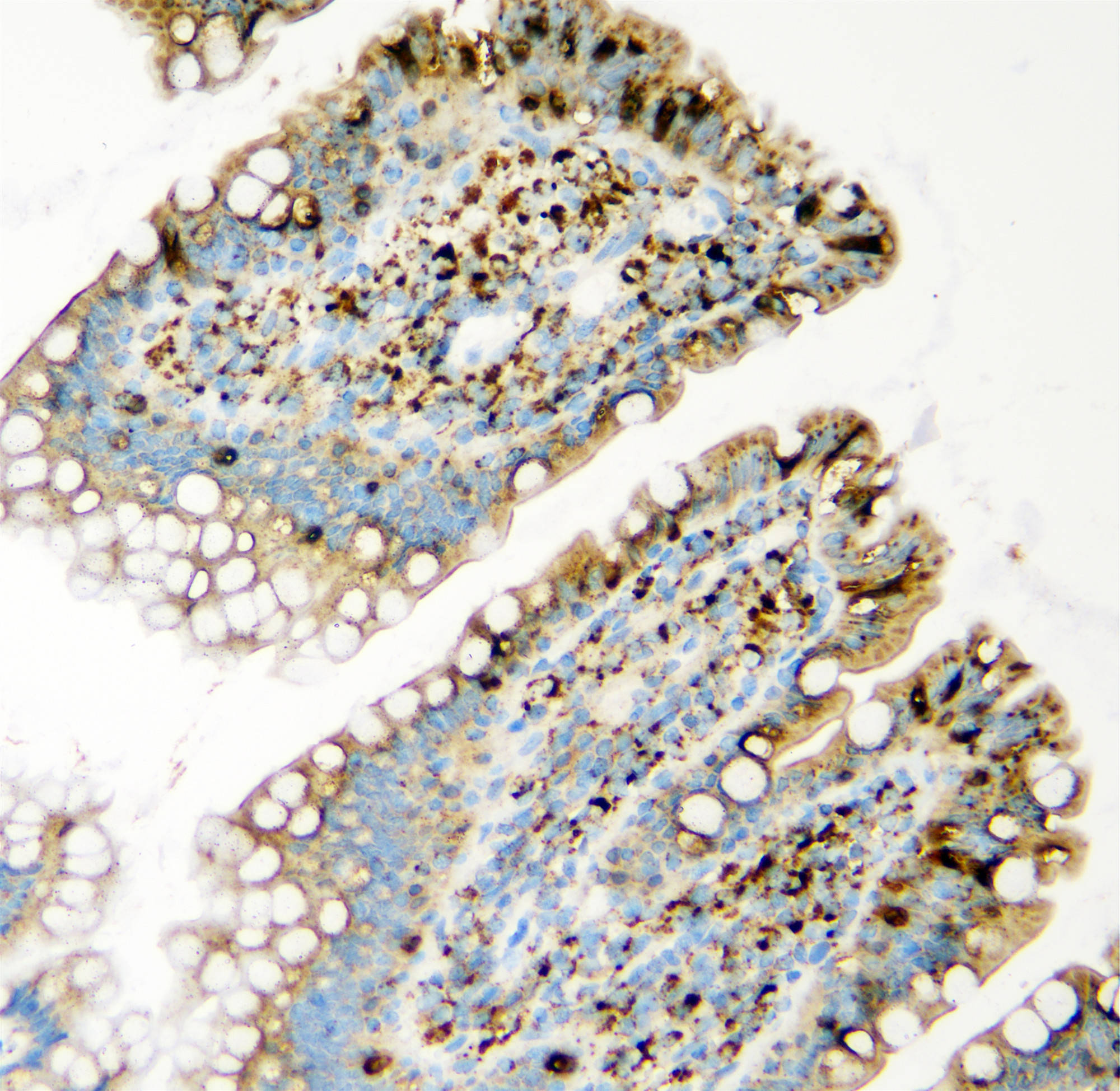
IHC analysis of Beta Galactosidase/GLB1 using anti-Beta Galactosidase/GLB1 antibody (A01829-3).
Beta Galactosidase/GLB1 was detected in a paraffin-embedded section of rat intestine tissue. Biotinylated goat anti-rabbit IgG was used as secondary antibody. The tissue section was incubated with rabbit anti-Beta Galactosidase/GLB1 Antibody (A01829-3) at a dilution of 1:200 and developed using Strepavidin-Biotin-Complex (SABC) (Catalog # SA1022) with DAB (Catalog # AR1027) as the chromogen.
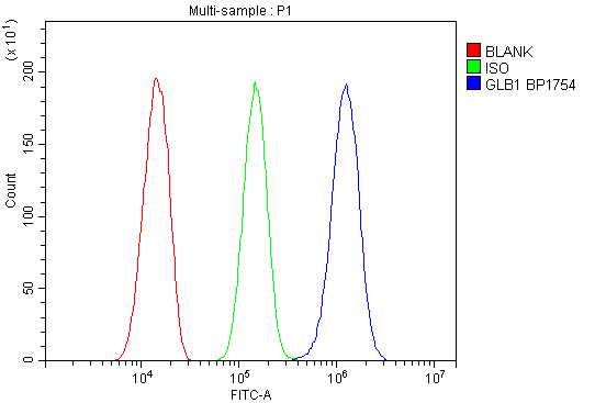
Flow Cytometry analysis of Hepa1-6 cells using anti-Beta Galactosidase/GLB1 antibody (A01829-3).
Overlay histogram showing Hepa1-6 cells stained with A01829-3 (Blue line). To facilitate intracellular staining, cells were fixed with 4% paraformaldehyde and permeabilized with permeabilization buffer. The cells were blocked with 10% normal goat serum. And then incubated with rabbit anti-Beta Galactosidase/GLB1 Antibody (A01829-3) at 1:100 dilution for 30 min at 20°C. Fluoro488 conjugated goat anti-rabbit IgG (BA1127) was used as secondary antibody at 1:100 dilution for 30 minutes at 20°C. Isotype control antibody (Green line) was rabbit IgG at 1:100 dilution used under the same conditions. Unlabelled sample without incubation with primary antibody and secondary antibody (Red line) was used as a blank control.
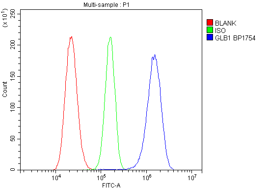
Flow Cytometry analysis of RH-35 cells using anti-Beta Galactosidase/GLB1 antibody (A01829-3).
Overlay histogram showing RH-35 cells stained with A01829-3 (Blue line). To facilitate intracellular staining, cells were fixed with 4% paraformaldehyde and permeabilized with permeabilization buffer. The cells were blocked with 10% normal goat serum. And then incubated with rabbit anti-Beta Galactosidase/GLB1 Antibody (A01829-3) at 1:100 dilution for 30 min at 20°C. Fluoro488 conjugated goat anti-rabbit IgG (BA1127) was used as secondary antibody at 1:100 dilution for 30 minutes at 20°C. Isotype control antibody (Green line) was rabbit IgG at 1:100 dilution used under the same conditions. Unlabelled sample without incubation with primary antibody and secondary antibody (Red line) was used as a blank control.
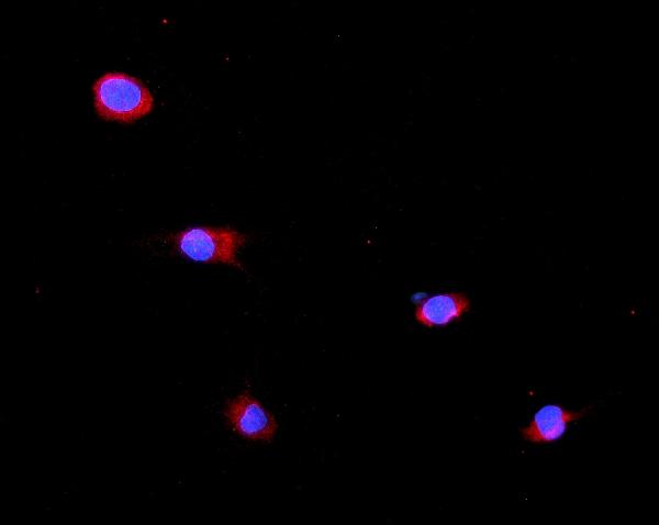
ICC/IF analysis of Beta Galactosidase/GLB1 using anti-Beta Galactosidase/GLB1 antibody (A01829-3).
Beta Galactosidase/GLB1 was detected in an immunocytochemical section of NRK cells. Fluoro550-conjugated Anti-rabbit IgG Secondary Antibody (red)(Catalog#BA1135) was used as secondary antibody. The section was counterstained with DAPI (Catalog # AR1176) (Blue).

| Western blot (WB): | 1:500-2000 |
| Immunohistochemistry (IHC): | 1:50-400 |
| Immunocytochemistry/Immunofluorescence (ICC/IF): | 1:50-400 |
| Flow Cytometry (Fixed): | 1:50-200 |
| Enzyme linked immunosorbent assay (ELISA): | 1:100-1000 |
| (Boiling the paraffin sections in 10mM citrate buffer,pH6.0,or PH8.0 EDTA repair liquid for 20 mins is required for the staining of formalin/paraffin sections.) Optimal working dilutions must be determined by end user. | |
Western blot analysis of Beta Galactosidase/GLB1 using anti-Beta Galactosidase/GLB1 antibody (A01829-3). The sample well of each lane was loaded with 30 ug of sample under reducing conditions.
Lane 1: rat lung tissue lysates,
Lane 2: rat liver tissue lysates,
Lane 3: rat testis tissue lysates,
Lane 4: mouse liver tissue lysates,
Lane 5: mouse testis tissue lysates,
Lane 6: mouse HEPA1-6 whole cell lysates,
Lane 7: mouse Neuro-2a whole cell lysates.
After electrophoresis, proteins were transferred to a membrane. Then the membrane was incubated with rabbit anti-Beta Galactosidase/GLB1 antigen affinity purified polyclonal antibody (A01829-3) at a dilution of 1:1000 and probed with a goat anti-rabbit IgG-HRP secondary antibody (Catalog # BA1054). The signal is developed using ECL Plus Western Blotting Substrate (Catalog # AR1197). A specific band was detected for Beta Galactosidase/GLB1 at approximately 65-85 kDa. The expected band size for Beta Galactosidase/GLB1 is at 73 kDa.
IHC analysis of Beta Galactosidase/GLB1 using anti-Beta Galactosidase/GLB1 antibody (A01829-3).
Beta Galactosidase/GLB1 was detected in a paraffin-embedded section of rat intestine tissue. Biotinylated goat anti-rabbit IgG was used as secondary antibody. The tissue section was incubated with rabbit anti-Beta Galactosidase/GLB1 Antibody (A01829-3) at a dilution of 1:200 and developed using Strepavidin-Biotin-Complex (SABC) (Catalog # SA1022) with DAB (Catalog # AR1027) as the chromogen.
Flow Cytometry analysis of Hepa1-6 cells using anti-Beta Galactosidase/GLB1 antibody (A01829-3).
Overlay histogram showing Hepa1-6 cells stained with A01829-3 (Blue line). To facilitate intracellular staining, cells were fixed with 4% paraformaldehyde and permeabilized with permeabilization buffer. The cells were blocked with 10% normal goat serum. And then incubated with rabbit anti-Beta Galactosidase/GLB1 Antibody (A01829-3) at 1:100 dilution for 30 min at 20°C. Fluoro488 conjugated goat anti-rabbit IgG (BA1127) was used as secondary antibody at 1:100 dilution for 30 minutes at 20°C. Isotype control antibody (Green line) was rabbit IgG at 1:100 dilution used under the same conditions. Unlabelled sample without incubation with primary antibody and secondary antibody (Red line) was used as a blank control.
Flow Cytometry analysis of RH-35 cells using anti-Beta Galactosidase/GLB1 antibody (A01829-3).
Overlay histogram showing RH-35 cells stained with A01829-3 (Blue line). To facilitate intracellular staining, cells were fixed with 4% paraformaldehyde and permeabilized with permeabilization buffer. The cells were blocked with 10% normal goat serum. And then incubated with rabbit anti-Beta Galactosidase/GLB1 Antibody (A01829-3) at 1:100 dilution for 30 min at 20°C. Fluoro488 conjugated goat anti-rabbit IgG (BA1127) was used as secondary antibody at 1:100 dilution for 30 minutes at 20°C. Isotype control antibody (Green line) was rabbit IgG at 1:100 dilution used under the same conditions. Unlabelled sample without incubation with primary antibody and secondary antibody (Red line) was used as a blank control.
ICC/IF analysis of Beta Galactosidase/GLB1 using anti-Beta Galactosidase/GLB1 antibody (A01829-3).
Beta Galactosidase/GLB1 was detected in an immunocytochemical section of NRK cells. Fluoro550-conjugated Anti-rabbit IgG Secondary Antibody (red)(Catalog#BA1135) was used as secondary antibody. The section was counterstained with DAPI (Catalog # AR1176) (Blue).
Western blot analysis of Beta Galactosidase/GLB1 using anti-Beta Galactosidase/GLB1 antibody (A01829-3). The sample well of each lane was loaded with 30 ug of sample under reducing conditions.
Lane 1: rat lung tissue lysates,
Lane 2: rat liver tissue lysates,
Lane 3: rat testis tissue lysates,
Lane 4: mouse liver tissue lysates,
Lane 5: mouse testis tissue lysates,
Lane 6: mouse HEPA1-6 whole cell lysates,
Lane 7: mouse Neuro-2a whole cell lysates.
After electrophoresis, proteins were transferred to a membrane. Then the membrane was incubated with rabbit anti-Beta Galactosidase/GLB1 antigen affinity purified polyclonal antibody (A01829-3) at a dilution of 1:1000 and probed with a goat anti-rabbit IgG-HRP secondary antibody (Catalog # BA1054). The signal is developed using ECL Plus Western Blotting Substrate (Catalog # AR1197). A specific band was detected for Beta Galactosidase/GLB1 at approximately 65-85 kDa. The expected band size for Beta Galactosidase/GLB1 is at 73 kDa.
IHC analysis of Beta Galactosidase/GLB1 using anti-Beta Galactosidase/GLB1 antibody (A01829-3).
Beta Galactosidase/GLB1 was detected in a paraffin-embedded section of rat intestine tissue. Biotinylated goat anti-rabbit IgG was used as secondary antibody. The tissue section was incubated with rabbit anti-Beta Galactosidase/GLB1 Antibody (A01829-3) at a dilution of 1:200 and developed using Strepavidin-Biotin-Complex (SABC) (Catalog # SA1022) with DAB (Catalog # AR1027) as the chromogen.
Flow Cytometry analysis of Hepa1-6 cells using anti-Beta Galactosidase/GLB1 antibody (A01829-3).
Overlay histogram showing Hepa1-6 cells stained with A01829-3 (Blue line). To facilitate intracellular staining, cells were fixed with 4% paraformaldehyde and permeabilized with permeabilization buffer. The cells were blocked with 10% normal goat serum. And then incubated with rabbit anti-Beta Galactosidase/GLB1 Antibody (A01829-3) at 1:100 dilution for 30 min at 20°C. Fluoro488 conjugated goat anti-rabbit IgG (BA1127) was used as secondary antibody at 1:100 dilution for 30 minutes at 20°C. Isotype control antibody (Green line) was rabbit IgG at 1:100 dilution used under the same conditions. Unlabelled sample without incubation with primary antibody and secondary antibody (Red line) was used as a blank control.
Flow Cytometry analysis of RH-35 cells using anti-Beta Galactosidase/GLB1 antibody (A01829-3).
Overlay histogram showing RH-35 cells stained with A01829-3 (Blue line). To facilitate intracellular staining, cells were fixed with 4% paraformaldehyde and permeabilized with permeabilization buffer. The cells were blocked with 10% normal goat serum. And then incubated with rabbit anti-Beta Galactosidase/GLB1 Antibody (A01829-3) at 1:100 dilution for 30 min at 20°C. Fluoro488 conjugated goat anti-rabbit IgG (BA1127) was used as secondary antibody at 1:100 dilution for 30 minutes at 20°C. Isotype control antibody (Green line) was rabbit IgG at 1:100 dilution used under the same conditions. Unlabelled sample without incubation with primary antibody and secondary antibody (Red line) was used as a blank control.
ICC/IF analysis of Beta Galactosidase/GLB1 using anti-Beta Galactosidase/GLB1 antibody (A01829-3).
Beta Galactosidase/GLB1 was detected in an immunocytochemical section of NRK cells. Fluoro550-conjugated Anti-rabbit IgG Secondary Antibody (red)(Catalog#BA1135) was used as secondary antibody. The section was counterstained with DAPI (Catalog # AR1176) (Blue).
 联系我们
联系我们027-67845390
 关注我们
关注我们
本司产品仅用于科研,不用于临床诊断和治疗
联系方式:027-67845390/1/2 技术支持:武汉丰网
© 1993-2025 Boster Biological Technology co.Itd E-mail:boster@boster.com
鄂ICP备05005548号-2
 鄂公网安备 42018502007312号

 积分商城
积分商城  购物车
购物车  登录/注册
登录/注册  您当前的位置:
 说明书
说明书 一键复制产品信息
 成功添加到购物车
 微信客服
微信客服


 微信扫一扫立即咨询
