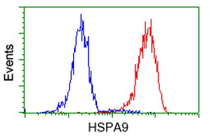
Flow cytometric Analysis of Hela cells, using anti-HSPA9 antibody, (Red), compared to a nonspecific negative control antibody, (Blue).
all(13)
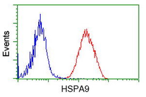
Flow cytometric Analysis of Jurkat cells, using anti-HSPA9 antibody, (Red), compared to a nonspecific negative control antibody, (Blue).
all(13) 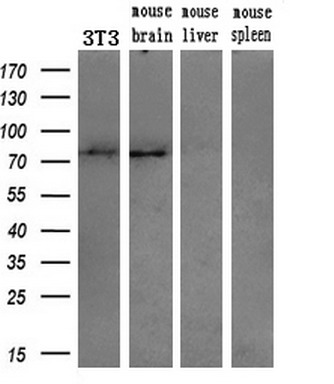
Western blot analysis of extracts (10ug) from a mouse cell line and 3 different mouse tissues by using anti-HSPA9 monoclonal antibody (1:200).
all(13) 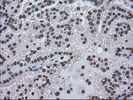
Immunohistochemical staining of paraffin-embedded Carcinoma of Human kidney tissue using anti-HSPA9 mouse monoclonal antibody. (Heat-induced epitope retrieval by 10mM citric buffer, pH6.0, 100°C for 10min, M02561-1)
all(13) 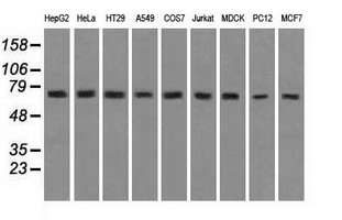
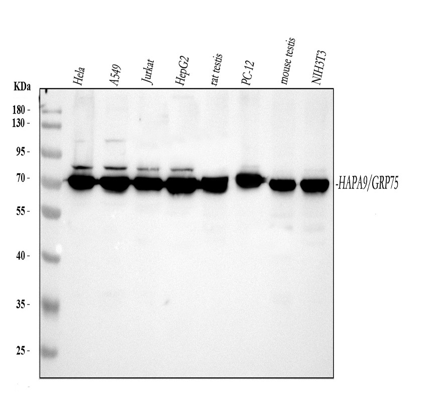
Western blot analysis of extracts (35ug) from 9 different cell lines by usin g anti-HSPA9 monoclonal antibody (HepG2: human; HeLa: human; SVT2: mouse; A549: human; COS7: monkey; Jurkat: human; MDCK: canine; PC12: rat; MCF7: human).
all(13) 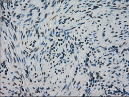
Immunohistochemical staining of paraffin-embedded Human endometrium tissue within the normal limits using anti-HSPA9 mouse monoclonal antibody. (Heat-induced epitope retrieval by 10mM citric buffer, pH6.0, 100°C for 10min, M02561-1)
all(13) 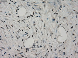
Immunohistochemical staining of paraffin-embedded Carcinoma of Human pancreas tissue using anti-HSPA9 mouse monoclonal antibody. (Heat-induced epitope retrieval by 10mM citric buffer, pH6.0, 100°C for 10min, M02561-1)
all(13) 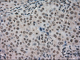
Immunohistochemical staining of paraffin-embedded Adenocarcinoma of Human breast tissue using anti-HSPA9 mouse monoclonal antibody. (Heat-induced epitope retrieval by 10mM citric buffer, pH6.0, 100°C for 10min, M02561-1)
all(13) 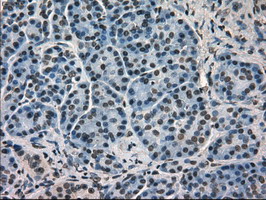
Immunohistochemical staining of paraffin-embedded Human pancreas tissue within the normal limits using anti-HSPA9 mouse monoclonal antibody. (Heat-induced epitope retrieval by 10mM citric buffer, pH6.0, 100°C for 10min, M02561-1)
all(13) 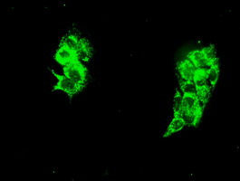
Immunofluorescent staining of HepG2 cells using anti-HSPA9 mouse monoclonal antibody.
all(13) 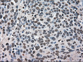
Immunohistochemical staining of paraffin-embedded Adenocarcinoma of Human ovary tissue using anti-HSPA9 mouse monoclonal antibody. (Heat-induced epitope retrieval by 10mM citric buffer, pH6.0, 100°C for 10min, M02561-1)
all(13) 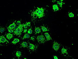
Anti-HSPA9 mouse monoclonal antibody immunofluorescent staining of COS7 cells transiently transfected by pCMV6-ENTRY HSPA9 .
all(13) 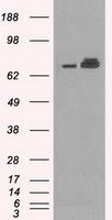
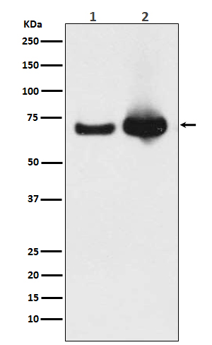
HEK293T cells were transfected with the pCMV6-ENTRY control (Left lane) or pCMV6-ENTRY HSPA9 (Right lane) cDNA for 48 hrs and lysed. Equivalent amounts of cell lysates (5 ug per lane) were separated by SDS-PAGE and immunoblotted with anti-HSPA9(Cat# M02561-1).
all(13) | Western blot (WB): | 1:1000~2000 |
| Immunohistochemistry (IHC): | 1:50 |
| Immunocytochemistry/Immunofluorescence (ICC/IF): | 1:50 |
| Flow cytometry (FCM): | 1:100 |
Flow cytometric Analysis of Hela cells, using anti-HSPA9 antibody, (Red), compared to a nonspecific negative control antibody, (Blue).
Flow cytometric Analysis of Jurkat cells, using anti-HSPA9 antibody, (Red), compared to a nonspecific negative control antibody, (Blue).
Western blot analysis of extracts (10ug) from a mouse cell line and 3 different mouse tissues by using anti-HSPA9 monoclonal antibody (1:200).
Immunohistochemical staining of paraffin-embedded Carcinoma of Human kidney tissue using anti-HSPA9 mouse monoclonal antibody. (Heat-induced epitope retrieval by 10mM citric buffer, pH6.0, 100°C for 10min, M02561-1)
Western blot analysis of extracts (35ug) from 9 different cell lines by usin g anti-HSPA9 monoclonal antibody (HepG2: human; HeLa: human; SVT2: mouse; A549: human; COS7: monkey; Jurkat: human; MDCK: canine; PC12: rat; MCF7: human).
Immunohistochemical staining of paraffin-embedded Human endometrium tissue within the normal limits using anti-HSPA9 mouse monoclonal antibody. (Heat-induced epitope retrieval by 10mM citric buffer, pH6.0, 100°C for 10min, M02561-1)
Immunohistochemical staining of paraffin-embedded Carcinoma of Human pancreas tissue using anti-HSPA9 mouse monoclonal antibody. (Heat-induced epitope retrieval by 10mM citric buffer, pH6.0, 100°C for 10min, M02561-1)
Immunohistochemical staining of paraffin-embedded Adenocarcinoma of Human breast tissue using anti-HSPA9 mouse monoclonal antibody. (Heat-induced epitope retrieval by 10mM citric buffer, pH6.0, 100°C for 10min, M02561-1)
Immunohistochemical staining of paraffin-embedded Human pancreas tissue within the normal limits using anti-HSPA9 mouse monoclonal antibody. (Heat-induced epitope retrieval by 10mM citric buffer, pH6.0, 100°C for 10min, M02561-1)
Immunofluorescent staining of HepG2 cells using anti-HSPA9 mouse monoclonal antibody.
Immunohistochemical staining of paraffin-embedded Adenocarcinoma of Human ovary tissue using anti-HSPA9 mouse monoclonal antibody. (Heat-induced epitope retrieval by 10mM citric buffer, pH6.0, 100°C for 10min, M02561-1)
Anti-HSPA9 mouse monoclonal antibody immunofluorescent staining of COS7 cells transiently transfected by pCMV6-ENTRY HSPA9 .
HEK293T cells were transfected with the pCMV6-ENTRY control (Left lane) or pCMV6-ENTRY HSPA9 (Right lane) cDNA for 48 hrs and lysed. Equivalent amounts of cell lysates (5 ug per lane) were separated by SDS-PAGE and immunoblotted with anti-HSPA9(Cat# M02561-1).
 联系我们
联系我们027-67845390
 关注我们
关注我们
本司产品仅用于科研,不用于临床诊断和治疗
联系方式:027-67845390/1/2 技术支持:武汉丰网
© 1993-2025 Boster Biological Technology co.Itd E-mail:boster@boster.com
鄂ICP备05005548号-2
 鄂公网安备 42018502007312号

 积分商城
积分商城  购物车
购物车  登录/注册
登录/注册  您当前的位置:
  说明书
说明书 一键复制产品信息
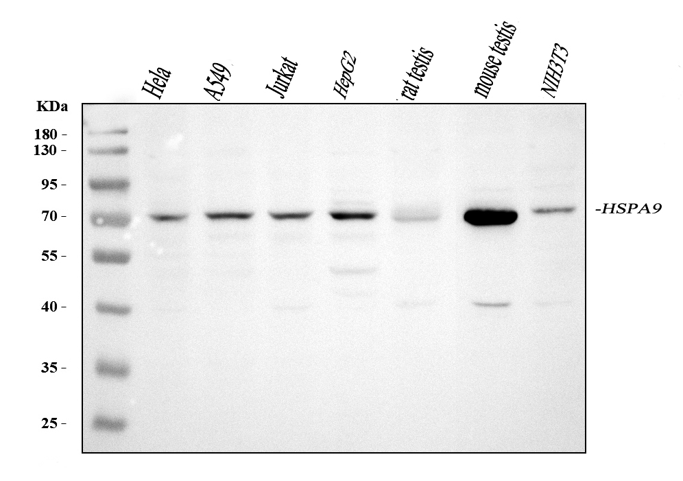
 成功添加到购物车
 微信客服
微信客服


 微信扫一扫立即咨询
