| Western blot (WB): | 1:500-2000 |
| Immunohistochemistry (IHC): | 1:50-400 |
| Immunocytochemistry/Immunofluorescence (ICC/IF): | 1:50-400 |
| Flow Cytometry (Fixed): | 1:50-200 |
| (Boiling the paraffin sections in 10mM citrate buffer,pH6.0,or PH8.0 EDTA repair liquid for 20 mins is required for the staining of formalin/paraffin sections.) Optimal working dilutions must be determined by end user. | |

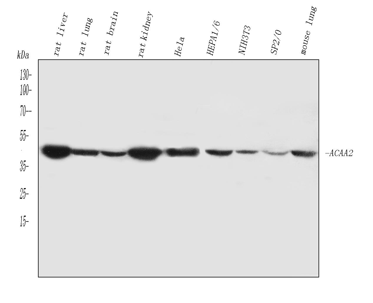
Western blot analysis of ACAA2(原PB10022PB1075) using anti-ACAA2(原PB10022PB1075) antibody (PB10022). The sample well of each lane was loaded with 30 ug of sample under reducing conditions.
Lane 1: rat liver tissue lysates,
Lane 2: rat lung tissue lysates,
Lane 3: rat brain tissue lysates,
Lane 4: rat kidney tissue lysates,
Lane 5: HELA whole cell lysates,
Lane 6: HEPA1/6 whole cell lysates,
Lane 7: NIH/3T3 whole cell lysates,
Lane 8: SP2/0 whole cell lysates,
Lane 9: mouse lung tissue lysates.
After electrophoresis, proteins were transferred to a membrane. Then the membrane was incubated with rabbit anti-ACAA2(原PB10022PB1075) antigen affinity purified polyclonal antibody (PB10022) at a dilution of 1:1000 and probed with a goat anti-rabbit IgG-HRP secondary antibody (Catalog # BA1054). The signal is developed using ECL Plus Western Blotting Substrate (Catalog # AR1197). A specific band was detected for ACAA2(原PB10022PB1075) at approximately 42 kDa. The expected band size for ACAA2(原PB10022PB1075) is at 42 kDa.

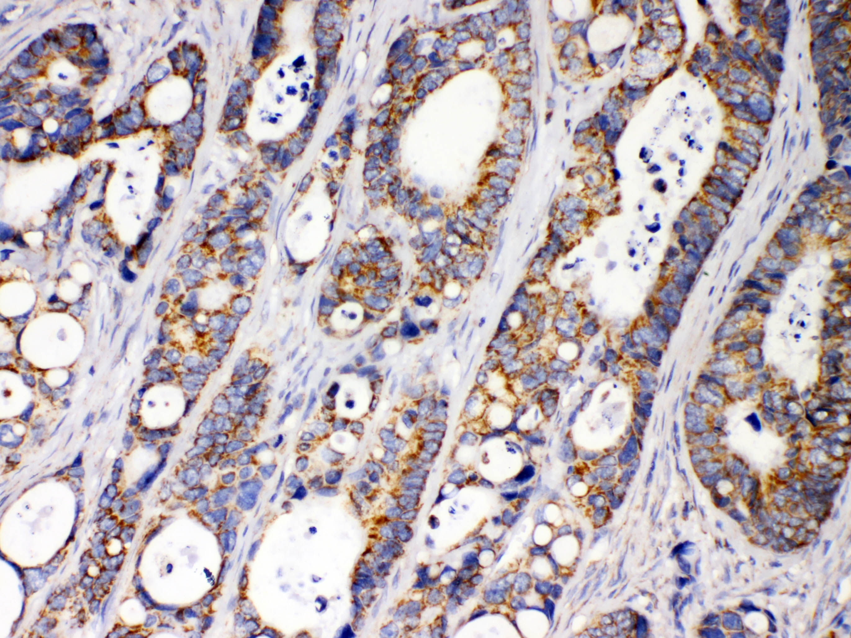
IHC analysis of ACAA2(原PB10022PB1075) using anti-ACAA2(原PB10022PB1075) antibody (PB10022).
ACAA2(原PB10022PB1075) was detected in a paraffin-embedded section of human intestinal cancer tissue. Biotinylated goat anti-rabbit IgG was used as secondary antibody. The tissue section was incubated with rabbit anti-ACAA2(原PB10022PB1075) Antibody (PB10022) at a dilution of 1:200 and developed using Strepavidin-Biotin-Complex (SABC) (Catalog # SA1022) with DAB (Catalog # AR1027) as the chromogen.

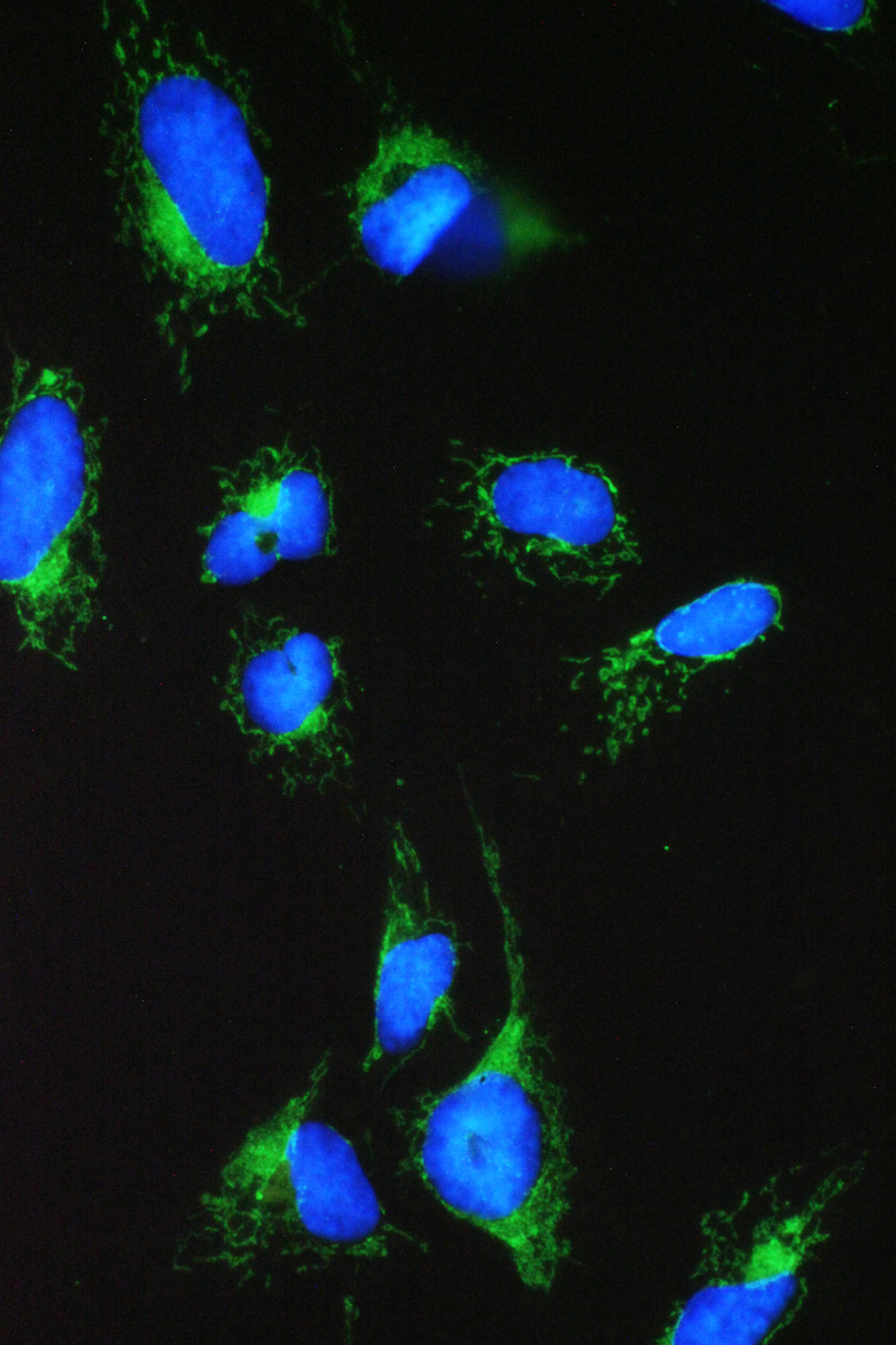
ICC/IF analysis of ACAA2 using anti- ACAA2 antibody (PB10022).
ACAA2 was detected in immunocytochemical section of U2OS cell. Enzyme antigen retrieval was performed using IHC enzyme antigen retrieval reagent (AR0022) for 15 mins. The cells were blocked with 10% goat serum. And then incubated with 2μg/mL rabbit anti- ACAA2 Antibody ((PB10022) overnight at 4°C. Fluoro488 Conjugated Goat Anti-Rabbit IgG (BA1127) was used as secondary antibody at 1:100 dilution and incubated for 30 minutes at 37°C. The section was counterstained with DAPI. Visualize using a fluorescence microscope and filter sets appropriate for the label used.

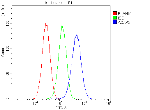
Flow Cytometry analysis of HepG2 cells using anti-ACAA2(原PB10022PB1075) antibody (PB10022).
Overlay histogram showing HepG2 cells stained with PB10022 (Blue line). To facilitate intracellular staining, cells were fixed with 4% paraformaldehyde and permeabilized with permeabilization buffer. The cells were blocked with 10% normal goat serum. And then incubated with rabbit anti-ACAA2(原PB10022PB1075) Antibody (PB10022) at 1:100 dilution for 30 min at 20°C. Fluoro488 conjugated goat anti-rabbit IgG (BA1127) was used as secondary antibody at 1:100 dilution for 30 minutes at 20°C. Isotype control antibody (Green line) was rabbit IgG at 1:100 dilution used under the same conditions. Unlabelled sample without incubation with primary antibody and secondary antibody (Red line) was used as a blank control.

Western blot analysis of ACAA2(原PB10022PB1075) using anti-ACAA2(原PB10022PB1075) antibody (PB10022). The sample well of each lane was loaded with 30 ug of sample under reducing conditions.
Lane 1: rat liver tissue lysates,
Lane 2: rat lung tissue lysates,
Lane 3: rat brain tissue lysates,
Lane 4: rat kidney tissue lysates,
Lane 5: HELA whole cell lysates,
Lane 6: HEPA1/6 whole cell lysates,
Lane 7: NIH/3T3 whole cell lysates,
Lane 8: SP2/0 whole cell lysates,
Lane 9: mouse lung tissue lysates.
After electrophoresis, proteins were transferred to a membrane. Then the membrane was incubated with rabbit anti-ACAA2(原PB10022PB1075) antigen affinity purified polyclonal antibody (PB10022) at a dilution of 1:1000 and probed with a goat anti-rabbit IgG-HRP secondary antibody (Catalog # BA1054). The signal is developed using ECL Plus Western Blotting Substrate (Catalog # AR1197). A specific band was detected for ACAA2(原PB10022PB1075) at approximately 42 kDa. The expected band size for ACAA2(原PB10022PB1075) is at 42 kDa.

IHC analysis of ACAA2(原PB10022PB1075) using anti-ACAA2(原PB10022PB1075) antibody (PB10022).
ACAA2(原PB10022PB1075) was detected in a paraffin-embedded section of human intestinal cancer tissue. Biotinylated goat anti-rabbit IgG was used as secondary antibody. The tissue section was incubated with rabbit anti-ACAA2(原PB10022PB1075) Antibody (PB10022) at a dilution of 1:200 and developed using Strepavidin-Biotin-Complex (SABC) (Catalog # SA1022) with DAB (Catalog # AR1027) as the chromogen.

ICC/IF analysis of ACAA2 using anti- ACAA2 antibody (PB10022).
ACAA2 was detected in immunocytochemical section of U2OS cell. Enzyme antigen retrieval was performed using IHC enzyme antigen retrieval reagent (AR0022) for 15 mins. The cells were blocked with 10% goat serum. And then incubated with 2μg/mL rabbit anti- ACAA2 Antibody ((PB10022) overnight at 4°C. Fluoro488 Conjugated Goat Anti-Rabbit IgG (BA1127) was used as secondary antibody at 1:100 dilution and incubated for 30 minutes at 37°C. The section was counterstained with DAPI. Visualize using a fluorescence microscope and filter sets appropriate for the label used.

Flow Cytometry analysis of HepG2 cells using anti-ACAA2(原PB10022PB1075) antibody (PB10022).
Overlay histogram showing HepG2 cells stained with PB10022 (Blue line). To facilitate intracellular staining, cells were fixed with 4% paraformaldehyde and permeabilized with permeabilization buffer. The cells were blocked with 10% normal goat serum. And then incubated with rabbit anti-ACAA2(原PB10022PB1075) Antibody (PB10022) at 1:100 dilution for 30 min at 20°C. Fluoro488 conjugated goat anti-rabbit IgG (BA1127) was used as secondary antibody at 1:100 dilution for 30 minutes at 20°C. Isotype control antibody (Green line) was rabbit IgG at 1:100 dilution used under the same conditions. Unlabelled sample without incubation with primary antibody and secondary antibody (Red line) was used as a blank control.


