-
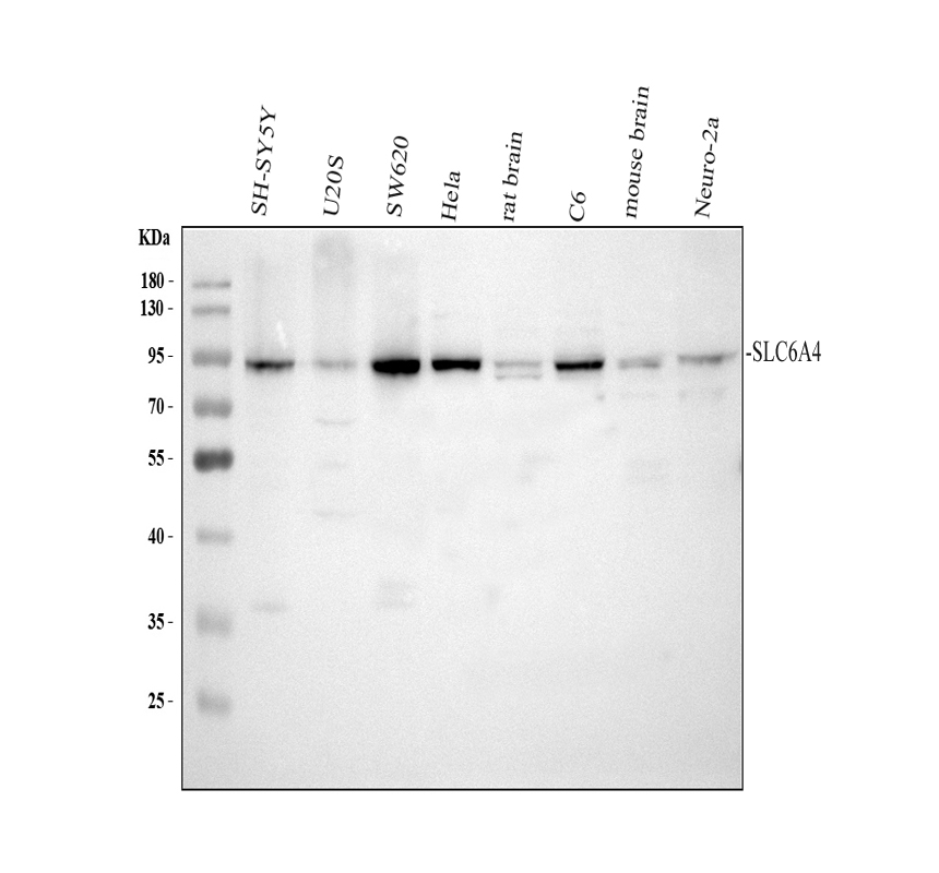
Western blot analysis of Serotonin transporter/SLC6A4 using anti-Serotonin transporter/SLC6A4 antibody (PB0442). The sample well of each lane was loaded with 30 ug of sample under reducing conditions.
Lane 1: human SH-SY5Y whole cell lysates,
Lane 2: human U2OS whole cell lysates,
Lane 3: human SW620 whole cell lysates,
Lane 4: human Hela whole cell lysates,
Lane 5: rat brain tissue lysates,
Lane 6: rat C6 whole cell lysates,
Lane 7: mouse brain tissue lysates,
Lane 8: mouse Neuro-2a whole cell lysates.
After electrophoresis, proteins were transferred to a membrane. Then the membrane was incubated with rabbit anti-Serotonin transporter/SLC6A4 antigen affinity purified polyclonal antibody (PB0442) at a dilution of 1:1000 and probed with a goat anti-rabbit IgG-HRP secondary antibody (Catalog # BA1054). The signal is developed using ECL Plus Western Blotting Substrate (Catalog # AR1197). A specific band was detected for Serotonin transporter/SLC6A4 at approximately 70-90 kDa. The expected band size for Serotonin transporter/SLC6A4 is at 70 kDa.
-
Western blot analysis of Serotonin transporter/SLC6A4 using anti-Serotonin transporter/SLC6A4 antibody (PB0442). The sample well of each lane was loaded with 30 ug of sample under reducing conditions.
Lane 1: human HepG2 whole cell lysates,
Lane 2: human Caco-2 whole cell lysates,
Lane 3: human A549 whole cell lysates,
Lane 4: human SH-SY5Y whole cell lysates.
After electrophoresis, proteins were transferred to a membrane. Then the membrane was incubated with rabbit anti-Serotonin transporter/SLC6A4 antigen affinity purified polyclonal antibody (PB0442) at a dilution of 1:1000 and probed with a goat anti-rabbit IgG-HRP secondary antibody (Catalog # BA1054). The signal is developed using ECL Plus Western Blotting Substrate (Catalog # AR1197). A specific band was detected for Serotonin transporter/SLC6A4 at approximately 70-90 kDa. The expected band size for Serotonin transporter/SLC6A4 is at 70 kDa.
-
Western blot analysis of Serotonin transporter/SLC6A4 using anti-Serotonin transporter/SLC6A4 antibody (PB0442). The sample well of each lane was loaded with 30 ug of sample under reducing conditions.
Lane 1: rat testis tissue lysates,
Lane 2: mouse testis tissue lysates.
After electrophoresis, proteins were transferred to a membrane. Then the membrane was incubated with rabbit anti-Serotonin transporter/SLC6A4 antigen affinity purified polyclonal antibody (PB0442) at a dilution of 1:1000 and probed with a goat anti-rabbit IgG-HRP secondary antibody (Catalog # BA1054). The signal is developed using ECL Plus Western Blotting Substrate (Catalog # AR1197). A specific band was detected for Serotonin transporter/SLC6A4 at approximately 70-90 kDa. The expected band size for Serotonin transporter/SLC6A4 is at 70 kDa.
-
IHC analysis of Serotonin transporter/SLC6A4 using anti-Serotonin transporter/SLC6A4 antibody (PB0442).
Serotonin transporter/SLC6A4 was detected in a paraffin-embedded section of mouse brain tissue. The tissue section was incubated with rabbit anti-Serotonin transporter/SLC6A4 Antibody (PB0442) at a dilution of 1:200 and developed using HRP Conjugated Rabbit IgG Super Vision Assay Kit (Catalog # SV0002) with DAB (Catalog # AR1027) as the chromogen.
-
IHC analysis of Serotonin transporter/SLC6A4 using anti-Serotonin transporter/SLC6A4 antibody (PB0442).
Serotonin transporter/SLC6A4 was detected in a paraffin-embedded section of rat brain tissue. The tissue section was incubated with rabbit anti-Serotonin transporter/SLC6A4 Antibody (PB0442) at a dilution of 1:200 and developed using HRP Conjugated Rabbit IgG Super Vision Assay Kit (Catalog # SV0002) with DAB (Catalog # AR1027) as the chromogen.
-
IF analysis of Serotonin transporter/SLC6A4 using anti-Serotonin transporter/SLC6A4 antibody (PB0442).
Serotonin transporter/SLC6A4 was detected in a paraffin-embedded section of mouse brain tissue. The tissue section was incubated with rabbit anti-Serotonin transporter/SLC6A4 Antibody (PB0442) at a dilution of 1:100. Cy3-conjugated Anti-rabbit IgG Secondary Antibody (red)(Catalog#BA1032) was used as secondary antibody. The section was counterstained with DAPI (Catalog # AR1176) (Blue).
-
IF analysis of Serotonin transporter/SLC6A4 using anti-Serotonin transporter/SLC6A4 antibody (PB0442).
Serotonin transporter/SLC6A4 was detected in a paraffin-embedded section of rat brain tissue. The tissue section was incubated with rabbit anti-Serotonin transporter/SLC6A4 Antibody (PB0442) at a dilution of 1:100. Cy3-conjugated Anti-rabbit IgG Secondary Antibody (red)(Catalog#BA1032) was used as secondary antibody. The section was counterstained with DAPI (Catalog # AR1176) (Blue).

 您当前的位置: 首页 > 产品列表
 联系我们
联系我们 关注我们
关注我们
 鄂公网安备 42018502007312号

 积分商城
积分商城  购物车
购物车  登录/注册
登录/注册 
 成功添加到购物车
 微信客服
微信客服


 微信扫一扫立即咨询
