欢迎访问博士德生物官网! 服务热线:027-67845390 |
您当前的位置: 首页 > 产品列表

筛选项

  • 检验物种

  • 应用范围

  • 宿主

  • 发货周期

  • 研究领域

  • 产品形态

共有 6 条产品信息

    • (3)

    Anti-APC Antibody

    货号:BA0645

    描述:Rabbit polyclonal antibody

    检验物种: human, mouse, rat

    应用范围: WB, IHC

    货期:现货

    说明书 文献数量(4) 一键复制产品信息

    加入购物车
  • Anti-APC Antibody

    筛选器: All WB IHC

    • Western blot analysis of APC using anti-APC antibody (BA0645). The sample well of each lane was loaded with 30 ug of sample under reducing conditions.
      Lane 1: Hela whole cell lysates,
      Lane 2: Colo320 whole cell lysates.
      After electrophoresis, proteins were transferred to a membrane. Then the membrane was incubated with rabbit anti-APC antigen affinity purified polyclonal antibody (BA0645) at a dilution of 1:1000 and probed with a goat anti-rabbit IgG-HRP secondary antibody (Catalog # BA1054). The signal is developed using ECL Plus Western Blotting Substrate (Catalog # AR1197). A specific band was detected for APC at approximately 160,312 kDa. The expected band size for APC is at 312 kDa.

    • IHC analysis of APC using anti-APC antibody (BA0645).
      APC was detected in a paraffin-embedded section of rat intestine tissue. Biotinylated goat anti-rabbit IgG was used as secondary antibody. The tissue section was incubated with rabbit anti-APC Antibody (BA0645) at a dilution of 1:200 and developed using Strepavidin-Biotin-Complex (SABC) (Catalog # SA1022) with DAB (Catalog # AR1027) as the chromogen.

    • IHC analysis of APC using anti-APC antibody (BA0645).
      APC was detected in a paraffin-embedded section of intestinal cancer tissue. Biotinylated goat anti-rabbit IgG was used as secondary antibody. The tissue section was incubated with rabbit anti-APC Antibody (BA0645) at a dilution of 1:200 and developed using Strepavidin-Biotin-Complex (SABC) (Catalog # SA1022) with DAB (Catalog # AR1027) as the chromogen.

    亮暗模式切换
    X
    • (5)

    Anti-AQP1 Antibody

    货号:BA0648

    描述:Rabbit polyclonal antibody

    检验物种: human, mouse, rat

    应用范围: WB, IHC, FCM

    货期:现货

    说明书 文献数量(8) 一键复制产品信息

    加入购物车
  • Anti-AQP1 Antibody

    筛选器: All WB IHC FCM

    • Western blot analysis of AQP1 using anti-AQP1 antibody (BA0648). The sample well of each lane was loaded with 30 ug of sample under reducing conditions.
      Lane 1: rat kidney tissue lysates,
      Lane 2: mouse kidney tissue lysates.
      After electrophoresis, proteins were transferred to a membrane. Then the membrane was incubated with rabbit anti-AQP1 antigen affinity purified polyclonal antibody (BA0648) at a dilution of 1:1000 and probed with a goat anti-rabbit IgG-HRP secondary antibody (Catalog # BA1054). The signal is developed using ECL Plus Western Blotting Substrate (Catalog # AR1197). A specific band was detected for AQP1 at approximately 25 kDa. The expected band size for AQP1 is at 29 kDa.

    • IHC analysis of AQP1 using anti-AQP1 antibody (BA0648) .
      AQP1 was detected in a paraffin-embedded section of human liver tissue. The tissue section was incubated with rabbit anti-AQP1 Antibody (BA0648) at a dilution of 1:200 and developed using HRP Conjugated Rabbit IgG Super Vision Assay Kit (Catalog # SV0002) with DAB (Catalog # AR1027) as the chromogen.

    • IHC analysis of AQP1 using anti-AQP1 antibody (BA0648) .
      AQP1 was detected in a paraffin-embedded section of mouse kidney tissue. The tissue section was incubated with rabbit anti-AQP1 Antibody (BA0648) at a dilution of 1:200 and developed using HRP Conjugated Rabbit IgG Super Vision Assay Kit (Catalog # SV0002) with DAB (Catalog # AR1027) as the chromogen.

    • IHC analysis of AQP1 using anti-AQP1 antibody (BA0648) .
      AQP1 was detected in a paraffin-embedded section of rat kidney tissue. The tissue section was incubated with rabbit anti-AQP1 Antibody (BA0648) at a dilution of 1:200 and developed using HRP Conjugated Rabbit IgG Super Vision Assay Kit (Catalog # SV0002) with DAB (Catalog # AR1027) as the chromogen.

    • Flow Cytometry analysis of Hela cells using anti-AQP1 antibody (BA0648).
      Overlay histogram showing Hela cells stained with BA0648 (Blue line). The cells were fixed with 4% paraformaldehyde and blocked with 10% normal goat serum. And then incubated with rabbit anti-AQP1 Antibody (BA0648) at 1:100 dilution for 30 min at 20°C. DyLight®488 conjugated goat anti-rabbit IgG (BA1127) was used as secondary antibody at 1:100 dilution for 30 minutes at 20°C. Isotype control antibody (Green line) was rabbit IgG at 1:100 dilution used under the same conditions. Unlabelled sample (Red line) was also used as a control.

    亮暗模式切换
    X
    • (3)

    Anti-AQP2 Antibody

    货号:BA0649

    描述:Rabbit polyclonal antibody

    检验物种: human, mouse, rat

    应用范围: IHC, WB

    货期:现货

    说明书 文献数量(1) 一键复制产品信息

    加入购物车
  • Anti-AQP2 Antibody

    筛选器: All IHC WB

    • IHC analysis of AQP2 using anti-AQP2 antibody (BA0649).
      AQP2 was detected in a paraffin-embedded section of rat kidney tissue. The tissue section was incubated with rabbit anti-AQP2 Antibody (BA0649) at a dilution of 1:200 and developed using HRP Conjugated Rabbit IgG Super Vision Assay Kit (Catalog # SV0002) with DAB (Catalog # AR1027) as the chromogen.

    • Western blot analysis of AQP2 using anti-AQP2 antibody (BA0649). The sample well of each lane was loaded with 30 ug of sample under reducing conditions.
      Lane 1: MCF-7 whole cell lysates,
      Lane 2: SW620 whole cell lysates,
      Lane 3: HT1080 whole cell lysates.
      After electrophoresis, proteins were transferred to a membrane. Then the membrane was incubated with rabbit anti-AQP2 antigen affinity purified polyclonal antibody (BA0649) at a dilution of 1:1000 and probed with a goat anti-rabbit IgG-HRP secondary antibody (Catalog # BA1054). The signal is developed using ECL Plus Western Blotting Substrate (Catalog # AR1197). A specific band was detected for AQP2 at approximately 26-37 kDa. The expected band size for AQP2 is at 29 kDa.

    • IHC analysis of AQP2 using anti-AQP2 antibody (BA0649).
      AQP2 was detected in frozen section of rat kidney tissue. The tissue section was incubated with rabbit anti-AQP2 Antibody (BA0649) at a dilution of 1:200 and developed using HRP Conjugated Rabbit IgG Super Vision Assay Kit (Catalog # SV0002) with DAB (Catalog # AR1027) as the chromogen.

    亮暗模式切换
    X
    • (4)

    Anti-Annexin A1/ANXA1 Antibody

    货号:BA0640

    描述:Rabbit polyclonal antibody

    检验物种: human, mouse, rat

    应用范围: WB, IHC, IP

    货期:现货

    说明书 文献数量(3) 一键复制产品信息

    加入购物车
  • Anti-Annexin A1/ANXA1 Antibody

    筛选器: All WB IHC IP

    • Western blot analysis of Annexin A1/ANXA1 using anti-Annexin A1/ANXA1 antibody (BA0640). The sample well of each lane was loaded with 30 ug of sample under reducing conditions.
      Lane 1: human Hela whole cell lysates,
      Lane 2: human A431 whole cell lysates,
      Lane 3: rat C6 whole cell lysates,
      Lane 4: mouse C2C12 whole cell lysates,
      Lane 5: mouse NIH/3T3 whole cell lysates.
      After electrophoresis, proteins were transferred to a membrane. Then the membrane was incubated with rabbit anti-Annexin A1/ANXA1 antigen affinity purified polyclonal antibody (BA0640) at a dilution of 1:1000 and probed with a goat anti-rabbit IgG-HRP secondary antibody (Catalog # BA1054). The signal is developed using ECL Plus Western Blotting Substrate (Catalog # AR1197). A specific band was detected for Annexin A1/ANXA1 at approximately 37 kDa. The expected band size for Annexin A1/ANXA1 is at 39 kDa.

    • IHC analysis of Annexin A1/ANXA1 using anti-Annexin A1/ANXA1 antibody (BA0640) .
      Annexin A1/ANXA1 was detected in a paraffin-embedded section of human colon cancer tissue. The tissue section was incubated with rabbit anti-Annexin A1/ANXA1 Antibody (BA0640) at a dilution of 1:200 and developed using HRP Conjugated Rabbit IgG Super Vision Assay Kit (Catalog # SV0002) with DAB (Catalog # AR1027) as the chromogen.

    • IHC analysis of Annexin A1/ANXA1 using anti-Annexin A1/ANXA1 antibody (BA0640) .
      Annexin A1/ANXA1 was detected in a paraffin-embedded section of human liver cancer tissue. The tissue section was incubated with rabbit anti-Annexin A1/ANXA1 Antibody (BA0640) at a dilution of 1:200 and developed using HRP Conjugated Rabbit IgG Super Vision Assay Kit (Catalog # SV0002) with DAB (Catalog # AR1027) as the chromogen.

    • IP analysis of Annexin A1/ANXA1 using anti-Annexin A1/ANXA1 antibody (BA0640) in A431 whole cell lysate.
      Western blot analysis of Annexin A1/ANXA1 using anti- Annexin A1/ANXA1 antibody (BA0640).
      Lane 1: A431 whole cell lysates(30ug),
      Lane 2: Rabbit control IgG instead of anti- Annexin A1/ANXA1 antibody in A431 whole cell lysate,
      Lane 3: anti- Annexin A1/ANXA1 antibody (2μg) + A431 whole cell lysate (500μg).
      After electrophoresis, proteins were transferred to a membrane. Then the membrane was incubated with rabbit anti- Annexin A1/ANXA1 antigen affinity purified polyclonal antibody (BA0640) at a dilution of 1:1000 and probed with a goat anti-rabbit IgG-HRP secondary antibody (Catalog # BA1054). The signal is developed using ECL Plus Western Blotting Substrate (Catalog # AR1197). A specific band was detected for Annexin A1/ANXA1 at approximately 37 kDa. The expected band size for Annexin A1/ANXA1 is at 39 kDa.

    亮暗模式切换
    X
    • (3)

    Anti-Annexin A2/ANXA2 Antibody

    货号:BA0641

    描述:Rabbit polyclonal antibody

    检验物种: human, mouse, rat

    应用范围: WB, IHC

    货期:现货

    说明书 文献数量(1) 一键复制产品信息

    加入购物车
  • Anti-Annexin A2/ANXA2 Antibody

    筛选器: All WB IHC

    • Western blot analysis of Annexin A2/ANXA2 using anti-Annexin A2/ANXA2 antibody (BA0641). The sample well of each lane was loaded with 30 ug of sample under reducing conditions.
      Lane 1: mouse spleen tissue lysates,
      Lane 2: mouse NIH/3T3 whole cell lysates.
      After electrophoresis, proteins were transferred to a membrane. Then the membrane was incubated with rabbit anti-Annexin A2/ANXA2 antigen affinity purified polyclonal antibody (BA0641) at a dilution of 1:1000 and probed with a goat anti-rabbit IgG-HRP secondary antibody (Catalog # BA1054). The signal is developed using ECL Plus Western Blotting Substrate (Catalog # AR1197). A specific band was detected for Annexin A2/ANXA2 at approximately 36 kDa. The expected band size for Annexin A2/ANXA2 is at 39 kDa.

    • IHC analysis of Annexin A2/ANXA2 using anti-Annexin A2/ANXA2 antibody (BA0641) .
      Annexin A2/ANXA2 was detected in a paraffin-embedded section of human intestinal tissue. The tissue section was incubated with rabbit anti-Annexin A2/ANXA2 Antibody (BA0641) at a dilution of 1:200 and developed using HRP Conjugated Rabbit IgG Super Vision Assay Kit (Catalog # SV0002) with DAB (Catalog # AR1027) as the chromogen.

    • Western blot analysis of Annexin A2/ANXA2 using anti-Annexin A2/ANXA2 antibody (BA0641). The sample well of each lane was loaded with 30 ug of sample under reducing conditions.
      Lane 1: rat testis tissue lysates,
      Lane 2: rat lung tissue lysates,
      Lane 3: rat ovary tissue lysates,
      Lane 4: human MCF-7 whole cell lysates,
      Lane 5: human SMMC whole cell lysates,
      Lane 6: human A549 whole cell lysates,
      Lane 7: human Jurkat whole cell lysates.
      After electrophoresis, proteins were transferred to a membrane. Then the membrane was incubated with rabbit anti-Annexin A2/ANXA2 antigen affinity purified polyclonal antibody (BA0641) at a dilution of 1:1000 and probed with a goat anti-rabbit IgG-HRP secondary antibody (Catalog # BA1054). The signal is developed using ECL Plus Western Blotting Substrate (Catalog # AR1197). A specific band was detected for Annexin A2/ANXA2 at approximately 36 kDa. The expected band size for Annexin A2/ANXA2 is at 39 kDa.

    亮暗模式切换
    X
    • (4)

    Anti-Annexin V/ANXA5 Antibody

    货号:BA0644

    描述:Rabbit polyclonal antibody

    检验物种: human, mouse, rat

    应用范围: WB, IHC, ICC/IF

    货期:现货

    说明书 一键复制产品信息

    加入购物车
  • Anti-Annexin V/ANXA5 Antibody

    筛选器: All WB IHC ICC/IF

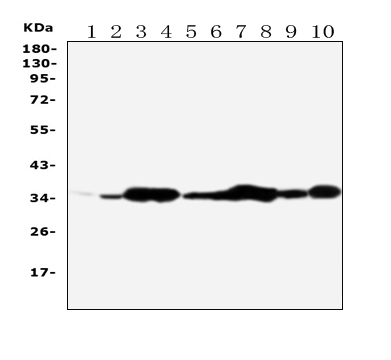
    • Western blot analysis of Annexin V/ANXA5 using anti-Annexin V/ANXA5 antibody (BA0644). The sample well of each lane was loaded with 30 ug of sample under reducing conditions.
      Lane 1: Rat brain tissue lysates,
      Lane 2: Rat skeletal muscle tissue lysates,
      Lane 3: Rat ovary tissue lysates,
      Lane 4: Rat lung tissue lysates,
      Lane 5: MCF-7 whole cell lysates,
      Lane 6: SMMC whole cell lysates,
      Lane 7: A549 whole cell lysates,
      Lane 8: JURKAT whole cell lysates,
      Lane 9: SGC whole cell lysates,
      Lane 10: HT1080 whole cell lysates.
      After electrophoresis, proteins were transferred to a membrane. Then the membrane was incubated with rabbit anti-Annexin V/ANXA5 antigen affinity purified polyclonal antibody (BA0644) at a dilution of 1:1000 and probed with a goat anti-rabbit IgG-HRP secondary antibody (Catalog # BA1054). The signal is developed using ECL Plus Western Blotting Substrate (Catalog # AR1197). A specific band was detected for Annexin V/ANXA5 at approximately 36 kDa. The expected band size for Annexin V/ANXA5 is at 36 kDa.

    • IHC analysis of Annexin V/ANXA5 using anti-Annexin V/ANXA5 antibody (BA0644).
      Annexin V/ANXA5 was detected in a paraffin-embedded section of rat liver tissue. Biotinylated goat anti-rabbit IgG was used as secondary antibody. The tissue section was incubated with rabbit anti-Annexin V/ANXA5 Antibody (BA0644) at a dilution of 1:200 and developed using Strepavidin-Biotin-Complex (SABC) (Catalog # SA1022) with DAB (Catalog # AR1027) as the chromogen.

    • ICC analysis of Annexin V/ANXA5 using anti- Annexin V/ANXA5 antibody (BA0644).
      Annexin V/ANXA5 was detected in an immunocytochemical section of HCT116 cells. The section was incubated with rabbit anti-Annexin V/ANXA5 Antibody (BA0644) at a dilution of 1:100. Biotinylated goat anti-rabbit IgG was used as secondary antibody. The section was developed using Strepavidin-Biotin-Complex (SABC)(Catalog # SA1022) with DAB (Catalog # AR1027) as the chromogen.

    • ICC analysis of Annexin V/ANXA5 using anti- Annexin V/ANXA5 antibody (BA0644).
      Annexin V/ANXA5 was detected in an immunocytochemical section of Hela cells. The section was incubated with rabbit anti-Annexin V/ANXA5 Antibody (BA0644) at a dilution of 1:100. Biotinylated goat anti-rabbit IgG was used as secondary antibody. The section was developed using Strepavidin-Biotin-Complex (SABC)(Catalog # SA1022) with DAB (Catalog # AR1027) as the chromogen.

    亮暗模式切换
    X

成功添加到购物车

查看购物车 继续购物

在线
客服

在线客服
服务时间:08:30 - 17:30

点击下列客服在线咨询:

服务热线

服务
热线

客服服务热线

027-67845390
客服服务热线

扫码

扫码
咨询

微信二维码 微信扫一扫立即咨询
返回顶部 顶部